This article has been reviewed according to Science X's editorial process and policies. Editors have highlighted the following attributes while ensuring the content's credibility:
fact-checked
trusted source
proofread
New excitation method of stimulated Raman scattering achieves natural-linewidth-limit spectral lines
 Schematics and (b) setup of Transient stimulated Raman scattering (T-SRS) spectroscopy. Credit: Qiaozhi Yu, Zhengjian Yao, Jiaqi Zhou, Wenhao Yu, Chenjie Zhuang, Yafeng Qi & Hanqing Xiong")
Stimulated Raman Scattering (SRS) has been developed as an essential quantitative contrast for chemical imaging in recent years. However, the spectral resolution of the mainstream SRS modalities is always lower than the state-of-the-art spontaneous Raman system.
This problem arises from the excitation strategy: the most widely used SRS modalities are all excited in the frequency domain. They have to compromise between the detection sensitivity and the spectral resolution: as the nonlinear process benefits from pulsed excitations, the fundamental time-energy uncertainty limits the spectral resolution.
In a new paper published in Light: Science & Applications, a team led by Dr. Hanqing Xiong from the National Biomedical Imaging Center, College of Future Technology at Peking University (Beijing, China) reported a novel method named transient stimulated Raman scattering (TSRS).
The team manipulated the interference of vibrational wave packets in the time domain by broadband femtosecond laser pulse trains and finally achieved natural-linewidth-limit Raman spectra at sub-mM sensitivity in a Fourier spectroscopic way. Furthermore, TSRS hyperspectral imaging of living Hela cells has been comprehensively performed in the Raman fingerprint region, the cell-silence region, and the popular C-H stretching region.
To show the advantage of the natural-linewidth-limit spectral resolution, The team also preliminarily constructed a set of high-density Raman probes with Raman mode intervals down to 12 cm-1 and further demonstrated its corresponding barcode imaging. The paper was published under the title of "Transient Stimulated Raman Scattering Spectroscopy and Imaging."
-
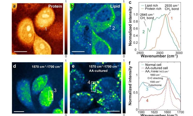
Label-free imaging of (a) proteins and (b) lipids of live cells. These two channels are unmixed from the overall hyperspectral data with the standard linear decomposition method. (c) Typical T-SRS spectra of proteins (region 1 in (a), red curve) and lipids (region 2 in (b), Green curve). (d) Typical T-SRS imaging of normal Hela cell and (e) 50-µM arachidonic acid (AA) cultured Hela cell in the [1570 cm-1, 1700 cm-1] range. (f) shows the spectra of the marked regions in (d) and (e), respectively. The red dashed curve is the C=C stretching mode Raman spectrum of pure AA. (g) Protein channel reference and (h) The C-D stretching band of d31- palmitic acid labeled cells. (i) T-SRS spectrum of the marked structure in (h). Scale bar: 10 μm for (a), (b), (d) and (e); 5 μm for (g) and (h). Credit: Qiaozhi Yu, Zhengjian Yao, Jiaqi Zhou, Wenhao Yu, Chenjie Zhuang, Yafeng Qi & Hanqing Xiong -
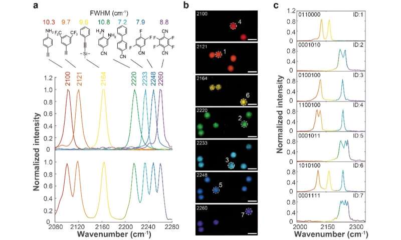
(a) Seven small-molecule probes and their corresponding T-SRS spectra in the triple-bond band. The upper panel shows the separated spectrum of each probe, the bottom panel shows the overall spectrum of the solution with seven probes mixed. (b) Signal of the encoded PMMA beads in each probe channel. (c) Spectra of typical encoded PMMA beads in the triple-bond band labeled in (b). Scale bar in (b): 10 μm. Credit: Qiaozhi Yu, Zhengjian Yao, Jiaqi Zhou, Wenhao Yu, Chenjie Zhuang, Yafeng Qi & Hanqing Xiong
Time-domain SRS techniques can find their origin in the 1980s, which is actually not new. However, previous time-domain SRS techniques cannot provide a sensitivity comparable to the widely used Frequency-domain methods. From the authors' point of view, the difference between the TSRS technique and other existing time-domain SRS methods is the use of stimulated Raman loss (SRL) as the signal.
SRL has a linear relation to the molecular concentration and the Raman cross-section, and it can be detected by classical heterodyne detection method to achieve the same shot-noise-limited sensitivity as the Frequency-domain methods. In order to construct a time-domain SRL signal, the authors abandoned the popular pump-probe excitation strategy.
Instead, they generated vibrational wave packet interference by two successive identical impulsive excitations with controlled time delay. The interference induces modulations on the SRL signal. A Fourier transform of the modulated SRL signal trace enables natural-linewidth-limit spectral lines.
"The spectral range of T-SRS imaging is only determined by the laser pulse bandwidths. The bandwidths of our excitation laser pulses can only support a spectral range of ~124 cm-1. We are constructing a laser system with much shorter pulses for TSRS, which may give full-range SRS spectra similar to the state-of-the-art spontaneous Raman system," said Dr. Xiong.
More information: Qiaozhi Yu et al, Transient stimulated Raman scattering spectroscopy and imaging, Light: Science & Applications (2024). DOI: 10.1038/s41377-024-01412-6
Provided by Chinese Academy of Sciences


