Improving deep optical microscope imaging of biological tissues
 Log scale Fourier spectra of speckles measured through anisotropic scattering samples according to different")
A team of researchers affiliated with UNIST has succeeded in developing a new optical microscope technology, capable of deeper imaging beyond the biological tissues. This breakthrough has been led by Professor Jung-Hoon Park and his research team in the Department of Biomedical Engineering at UNIST.
Optical imaging technology has emerged as an essential research tool for biomedical studies due to its high resolution and good tomography capability. However, the limited penetration depth of the optical microscope makes it difficult to observe biological tissues of more than 100 μm thickness. This is because strong light scattering, caused by various components of biological tissues, notably lipids and proteins, makes the subject out of focus, which then causes image blurring.
In this study, the research team showed that for wavefront shaping in thin anisotropic scattering media, such as biological tissues, they can optimize the wavefront shaping quality by simply limiting the numerical aperture (NA) of the incident wavefront.
In addition, using the same number of controlled modes, and therefore the same wavefront measurement time, the research team demonstrated that the wavefront shaped focus peak to background ratio can be increased by a factor of 2.1 while the energy delivery throughput can be increased by a factor of 8.9 through 710 μm thick brain tissue by just limiting the incident NA.
The research team anticipates that the new approach can open new avenues in a variety of biomedical applications where energy delivery enhancement or high-resolution imaging/photostimulation is required in a limited decorrelation time window or in light-starved environments.
-
Professor Jung-Hoon Park and his research team in the Department of Biomedical Engineering at UNIST. Credit: UNIST -
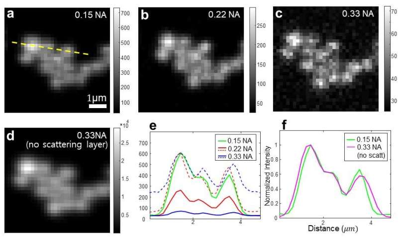
Laser scanning microscope image of 1 μm fluorescent beads through 710 μm thickness of brain slice by wavefront shaping. Credit: UNIST
More information: Hyungwon Jin et al, Limiting the incident NA for efficient wavefront shaping through thin anisotropic scattering media, Optica (2021). DOI: 10.1364/OPTICA.413174
Journal information: Optica


