New tool allows scientists to visualize 'nanoscale' processes

Chemists at UC San Diego have developed a new tool that allows scientists for the first time to see, at the scale of five billionths of a meter, "nanoscale" mixing processes occurring in liquids.
"Being able to look at nanoscale chemical gradients and reactions as they take place is just such a fundamental tool in biology, chemistry and all of material science," said Nathan Gianneschi, a professor of chemistry and biochemistry who headed the team that detailed the development in a paper in this week's issue of the journal Microscopy and Microanalysis. "With this new tool, we'll be able to look at the kinetics and dynamics of chemical interactions that we've never been able to see before."
Scientists have long relied on Transmission Electron Microscopy, or TEM, to see structures at the nanoscale. But that technique can take only static images and the subjects must be dried, or frozen and mounted within a vacuum chamber in order to be seen. As a result, researchers have been unable to view living processes or chemical reactions at the nanoscale, such as the growth and contraction within living cells of tiny fibers or nanoscale protrusions, essential in cell movement and division, or the changes caused by a chemical reaction in a liquid.
"As chemists, we could only really analyze the end products or bulk solution changes, or image at low resolution because we could never see events directly occur at the nanoscale," said Gianneschi.
Recent developments in Liquid Cell TEM, or LCTEM, have allowed scientists to finally take videos of nanoscale objects in liquids. But that technique has been limited by the inability to control the mixing of solutions, a requirement when trying to view and analyze the impact of a drug on a living cell or the reaction of two chemicals.
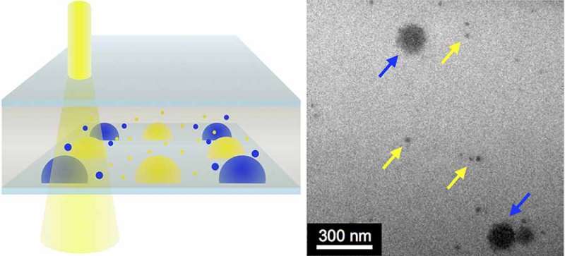
Joseph Patterson, a postdoctoral researcher in Gianneschi laboratory, working with researchers at SCIENION AG in Germany and Pacific Northwest National Laboratory, has taken a big step to resolving that problem by developing a technique as well as a tool that allows scientists to deposit tiny amounts of liquid—about 50 trillionths of a liter—within the viewing area of the LCTEM microscope.
"With this technique, we can view multiple components mixed together at the nanoscale within liquids, so, for example, one could look at biological materials and perhaps see how they respond to a drug," said Gianneschi. "That was never possible before."
"The benefits to basic research are huge," he added. "We will now be able to directly see the growth at the nanoscale of all kinds of things, like natural fibers or microtubules. There's a lot of interest on the part of researchers in understanding how the surfaces of nanoparticles affect chemical reactions or how nanoscale defects on the surfaces of materials develop. We can finally look at the interfaces on nanostructures so that we can optimize the development of new kinds of catalysts, paints and suspensions."
While the scientists have not yet used their tool to view chemical reactions in solution, they have demonstrated that the technique works to provide mixing using combinations of gold nanoparticles and other nanoscale crystals suspended in a liquid.
"What we've demonstrated is the proof of concept," said Gianneschi. "But that's what we'll be doing next."
Although this new tool won't allow scientists to actually view molecules in solution, Gianneschi said they should be able to see the impact of chemical reactions that are occurring on materials that are bigger than five nanometers, or five billionths of a meter.
"We won't be observing molecules colliding, but we will be able to observe single particles and collections of them, on the nanometer length scale," he added. "Observing these kinds of processes has been one of the key challenges in the field of nanoscience."
Provided by University of California - San Diego


