Page 12: Research news on Medical imaging

Medical imaging is a set of diagnostic techniques that noninvasively visualize internal anatomical structures and physiological processes using diverse physical modalities. Core methods include ionizing-radiation–based techniques (e.g., radiography, computed tomography, nuclear medicine, PET/SPECT), non-ionizing modalities (e.g., MRI, ultrasound, optical imaging), and hybrid systems (e.g., PET/CT, PET/MRI). These techniques exploit contrasts in tissue density, proton properties, acoustic impedance, or radiotracer distribution to generate qualitative and quantitative data. Medical imaging underpins clinical decision-making, image-guided interventions, treatment planning, and longitudinal monitoring, and increasingly incorporates advanced reconstruction algorithms, quantitative biomarkers, and AI-driven image analysis.

New method achieves 4D imaging of fluids in pores

A method based on CT (computed tomography)—a type of imaging that is widely used in hospitals—can help improve our understanding of CO2 storage, batteries, and processes in the body such as nutrient uptake.

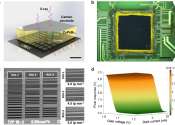
An ultracompact multimode meta-microscope

Versatility and miniaturization of imaging systems are of great importance in today's information society. Microscopic imaging techniques have always been indispensable for scientific research and disease diagnosis in the ...

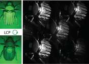
Enhanced 3D chemical imaging with phase-modulation

Understanding complex biological and biomedical systems is greatly aided by 3D imaging, which provides much more detailed information than traditional two-dimensional methods. However, live cell and tissue imaging remain ...

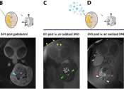
A new way to visualize brain cancer at the nanoscale level

Researchers from Brigham and Women's Hospital and the Massachusetts Institute of Technology (MIT) have unveiled unprecedentedly detailed images of brain cancer tissue through the use of a new microscopy technology called ...

page 12 from 16