Setting standards for protein localisation

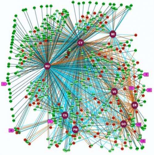
(Phys.org) —In order to understand how cells work, scientists first need to establish where every single protein in the cell resides. In the largest study of its kind to date, the two most widely used microscopy-based methods that can be applied to this task have been compared.
The international collaboration led by Conway Fellow, Professor Jeremy Simpson, UCD School of Biology & Environmental Science and Professor Emma Lundberg, KTH-Stockholm looked at more than 500 proteins using antibody-based localisation and fluorescent protein tagging methods.
Their findings show that by following a defined set of rules, both antibody-based and fluorescent-tagging methods are highly complementary to each other, and show a high degree of correlation for the determination of protein localisation in cells.
The study also provides a significant and experimentally validated data set of protein localisation in mammalian cells, which in itself is a valuable resource for the scientific community.
According to Professor Simpson, "The debate of whether antibodies or fluorescent protein tagging is the most reliable method to determine protein localisation has been going on for more than 15 years.
In addition to determining the localisation of more than 500 proteins, half of which had no previous localisation annotation, this study has allowed us to address a key experimental issue.
This work is expected to set the standard methodology for ultimately determining the localisation of the entire human proteome.
More information: Stadler, C. et al. Immunofluorescence and fluorescent-protein tagging show high correlation for protein localization in mammalian cells, Nature Methods (2013) Feb 24 doi: 10.1038/nmeth.2377. [Epub ahead of print].
Journal information: Nature Methods
Provided by University College Dublin


