Software visualizes the heart for doctors

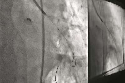
Siemens developed a procedure for angioplasty which supports doctors to make interventions at the heart faster and more precise. With angioplasty doctors open up an artery, potentially staving off more invasive procedures like heart bypass surgery. This method positions and expands a balloon, enclosed in a stent a metal stent, to open up a region in a coronary artery that can clog. To deploy the stent, doctors follow its position by X-ray fluoroscopy, but the procedure is limited by poor visibility and difficult use. This can be helped by use of intravascular ultrasound (IVUS) which implies the use of costly catheters.
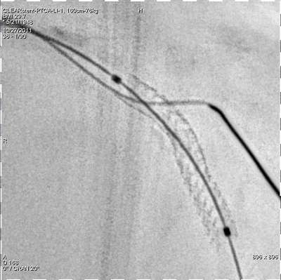
The visualizing tool CLEARstent makes this procedure easier. The software is for several angiography devices from Siemens which all work with low doses of radiation. After a few seconds imaging acquisition time, or using previously acquired images, CLEARstentcan tell clinicians if the stent is properly positioned and expanded. Doctors need to figure out where they want to place the stent and afterwards whether the stent is fully embedded in the blood vessel's wall. CLEARstent enables both these processes.
Experts from Siemens Healthcare and the global Siemens research organization Corporate Technology (CT) in Princeton, New Jersey, developed the software. CT researchers developed also the software CLEARstent Live which improves visibility, providing better images during live angioplasty when the heart is beating. Imaging of the stent by traditional X-ray fluoroscopy is complicated by the motion induced by the pumping heart. X-ray fluoroscopy tends to pick up blurry, faint images of the stent.

To improve the visibility of the stent, the CT team developed a robust method that detects the balloon markers at the stent's ends by a learning step with early snapshots of the stent. This preparative step would help the program identify the stent during live image acquisitions. To reduce blurry images further, CLEARstent Live performs a common method known as image registration. This method deforms and aligns the clearly defined stents in adjacent frames based on the marker position, thereby stabilizing the stent during real-time imaging. Another benefit for the doctor is that CLEARstent Live can overlay the images of the stent with those of the surrounding blood vessels. This ensures that live angioplasty is performed under the most physiological condition.


