Langerhans cells migrate to their final destination in multiple waves at different stages of embryonic development

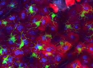
As our primary interface with the outside world, the skin needs to be able to protect itself against infectious threats. Specialized cells known as Langerhans cells (LCs) (see image) are an essential component of this defense, helping other immune cells to distinguish friend from foe. "These cells play an important role in maintaining tolerance to cutaneous antigen, while simultaneously promoting immune responses against any invading pathogens," explains Florent Ginhoux at the A*STAR Singapore Immunology Network.
LCs are the skin-based counterparts of dendritic cells (DCs), which reside in all tissues. However, LCs and tissue DCs are generated via distinct developmental pathways, and new research from Ginhoux and co-workers has uncovered unexpected complexity in the process by which the adult LC population arises over time.
Unlike DCs, which are generated continuously from bone marrow progenitors prior to their entry in the bloodstream and tissues, LCs are instead replenished by local precursors in the skin. In this sense, LCs are similar to microglia, immune cells residing exclusively in the brain, and Ginhoux suspected the two otherwise-similar cell types might arise from a common embryonic source. "We had previously shown that microglia derive from yolk sac progenitors," he says. "We hypothesized that LCs could be derived from yolk sac progenitors as well."
To test this model, the researchers used a genetic labeling strategy to trace the development of different cells in mouse embryos. Indeed, they confirmed that LCs are generated by precursors in the yolk sac, which migrate to the skin around the tenth day of mouse embryonic development, a process that requires 20 days in full. However, they were surprised to find that these yolk sac-derived cells form less than 10% of the adult LC population.
In fact, further lineage-tracing experiments revealed a later, larger wave of LC formation in the fetal liver, producing cells that migrate to the skin between the thirteenth and seventeenth day of embryonic development. "It was unexpected to find that the fetal liver contribution superseded that of the yolk sac," says Ginhoux. He notes that although both microglia and LCs appear to emerge from common progenitors in the yolk sac, the subsequent formation of the blood-brain barrier would most likely prevent equivalent migration of liver-derived microglial precursors to the brain.
This two-wave developmental process thus appears to be specifically limited to LCs, and Ginhoux and co-workers now hope to determine what, if any, functional value is gained by this stepwise developmental process.
More information: Hoeffel, G., Wang, Y., Greter, M., See, P., Teo, P., et al. Adult Langerhans cells derive predominantly from embryonic fetal liver monocytes with a minor contribution of yolk sac–derived macrophages. Journal of Experimental Medicine 209, 1167–1181 (2012). Article


