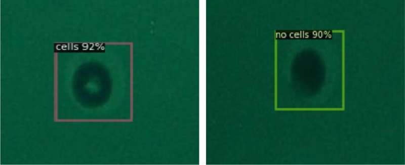
Presence of single cell (left) and absence of single cell (right). Credit: Toyohashi University Of Technology
A research team, led by Professor Moeto Nagai and comprised of researchers from the Department of Mechanical Engineering and the Electronic Inspired Interdisciplinary Research Institute (EIIRIS), Toyohashi University of Technology, has successfully used AI to achieve single-cell isolation.
The method involves using microwells to isolate single cells and then applying deep learning to the microscopic images containing single cells in the microwells. The machine learning model prepared by the team makes it possible to automatically detect single cells in microscopic images and reduce human effort. The acquisition of a large volume of single-cell data allows researchers to efficiently investigate the characteristics and functions of individual cells, which can lead to the establishment of new treatment methods.
A cell is the most basic unit of life, and elucidation of cell characteristics can contribute to a better understanding of diseased cells and thus to the development of new treatment methods. There has been a growing interest in developing methods of isolating single cells to study their functions. However, the physical isolation of single cells requires cell patterning tools. In addition, as the detection of single cells often relies on human eyes and classification, human effort has been a bottleneck that has hindered the acquisition of data.
Initially, the research team developed a method of isolating and trapping single cells in microwells of 30 μm diameter formed in a hydrogel patterned by optical patterning. The micro-patterned hydrogel offers the advantages of convenience and stability. These microwell structures allowed the research team to successfully isolate human single cells from a cell suspension. As hydrogel is highly biocompatible and can withstand a long incubation time in a cell medium, it was possible to extend the observation period of cell behavior.
Next, the research team classified the cells trapped in microwells of 30 μm diameter in terms of the presence or absence of single cells and used the images as training data to perform deep learning. The object detection model resulting from the learning could then be used to detect the presence of single cells based on input images. This made it possible to predict the presence of single cells with a mean Average Precision (mAP) of 0.989 (the higher the better) and an average inference time of 0.06 seconds (the shorter the better). This algorithm offers high detection accuracy while reducing experiment time, which leads to improving high-throughput single-cell analysis.
At first, the research team used bright-field microscopy images as input datasets, which are commonly used for the observation of single cells, but as these images did not offer high contrast, there were limits to the improvement of the detection capability. Performance stagnated at an mAP of 0.801 and an average inference time of 0.09 seconds.
The team then switched to cells stained with fluorescent dyes and the use of fluorescence microscopy images as the input datasets, which allowed them to achieve convergence at an mAP of 0.989 after 1,200 epochs of training. This indicates that preparing high-contrast input datasets, which make it easier for humans to detect the presence of the cells, is important also for the use of AI.
Research team leader Moeto Nagai explains, "We wanted to apply AI to the detection of single cells. As I had been performing mostly experiment-based research, the use of experimental data for AI research seemed to me to be a significant obstacle. However, the participation of graduate student Tanmay Debnath, the lead author of our study, who has experience in the research and development of AI technology, meant that we could rapidly make use of AI and ultimately led to the success of our development."
The single-cell isolation and detection developed by this research can also be used to automatically monitor the activities of single cells. This research achieves accurate and highly reliable automated cell detection, while reducing human labor. Future applications for single-cell analysis include medical engineering applications in a wide range of areas such as cancer diagnosis, immune response, and drug discovery screening, which will contribute to the discovery of new treatment methods.
The findings are published in the journal Scientific Reports.
More information: Tanmay Debnath et al, Automated detection of patterned single-cells within hydrogel using deep learning, Scientific Reports (2022). DOI: 10.1038/s41598-022-22774-0
Journal information: Scientific Reports
Provided by Toyohashi University of Technology


