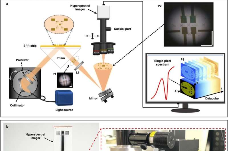
HSPRM system constructed in our laboratory. a Schematic diagram of the HSPRM system. P1 is a blurred SPR image contained in the reflected collimated beam. L1 is an achromatic imaging lens (NA = 0.4). P2 is a large FOV, clear SPR image formed by L1. P3 is a hyperspectral datacube for the selected area in P2. Scale bar = 2 mm. b Photograph of the laboratory-made HSPRM system. C1 and C2 are x-axis and y-axis precision moving stages used for selecting the region of interest in P2. C3 is a rotating holder mounted with three objectives of different magnifications. Credit: Nature Communications (2022). DOI: 10.1038/s41467-022-34196-7
Hyperspectral surface plasmon resonance microscopy (HSPRM) is an advanced analytical technique for spectral imaging and chemical and biological sensing, which enables high-resolution visualization and precise quantification of chemical and biological analytes.
A study published in Nature Communications describes a flexible HSPRM system that operates by using a hyperspectral microscope to analyze the selected area of an SPR image produced by a prism-based spectral SPR sensor.
The HSPRM system is developed by a research team from the Aerospace Information Research Institute (AIR) of the Chinese Academy of Sciences (CAS).
The HSPRM system enables monochromatic and polychromatic SPR imaging and single-pixel spectral SPR sensing, as well as two-dimensional quantification of thin films with measured resonance-wavelength images. It can measure SPR radiance spectra instead of conventional intensity spectra to improve the figure of merit (FOM) of single-pixel spectral SPR sensors and can also quantify two-dimensional profiles of thickness and refractive index for thin films by using measured resonance-wavelength images.
Pixel-by-pixel calibration of the incident beam collimation deviation was performed to remove pixel-to-pixel differences in SPR sensitivity.
The HSPRM system has a wide spectral range from 400 nm to 1,000 nm, an optional field of view from 0.884 mm2 to 0.003 mm2 and a high lateral resolution of 1.2μm.
Typical applications of the HSPRM system include quantification of single-layer graphene thickness distribution, in situ detection of inhomogeneous protein adsorption, and label-free single cell analysis.
More information: Ziwei Liu et al, Flexible hyperspectral surface plasmon resonance microscopy, Nature Communications (2022). DOI: 10.1038/s41467-022-34196-7
Journal information: Nature Communications
Provided by Chinese Academy of Sciences


