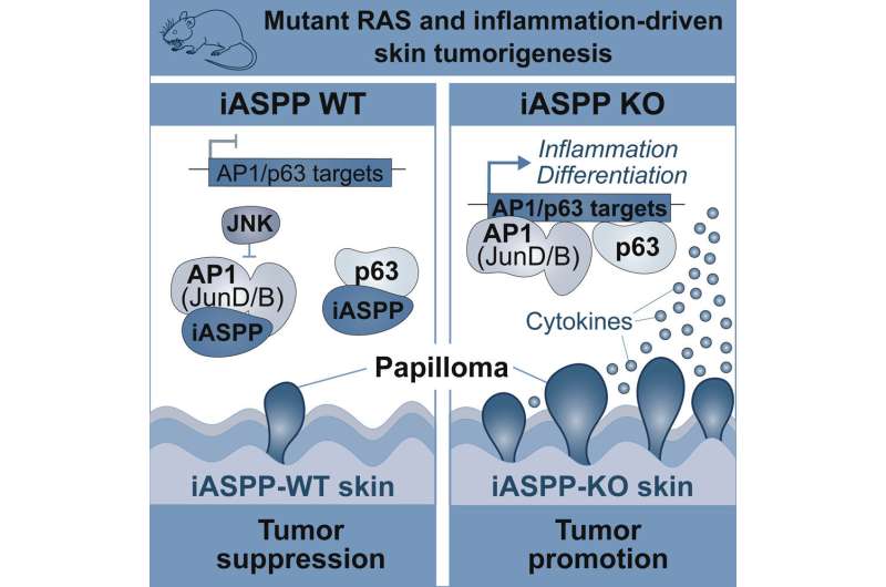
Graphical abstract. Credit: Cell Reports (2022). DOI: 10.1016/j.celrep.2022.111503
There is a tightly regulated balance between cell growth and death in healthy tissue. The loss of this balance is essentially what initiates cancer.
At a molecular level, that imbalance stems from a cell's acquisition of both activating mutations that promote its proliferation and the loss of inhibitory factors that suppress unregulated growth. In many cases, activating mutations in the RAS signaling pathway provide the former impetus, while mutations of the p53 tumor suppressor gene furnish the latter. Cancers driven by RAS signaling that are also associated with inflammation can, however, bypass the need for p53 pathway dysfunction. How they did this was not entirely clear.
To find out, Ludwig Oxford's Khatoun Al Moussawi, Kathryn Chung, Thomas Carroll and Artem Smirnov in Branch Director Xin Lu's laboratory and colleagues at Goethe University in Germany explored carcinogenesis in a well-established mouse model of mutant RAS- and inflammation-driven skin cancer. Their findings, described in Cell Reports, reveal that iASPP, which typically promotes tumor growth by inhibiting p53, unexpectedly suppresses cancer initiation in this context.
This, they report, is because iASPP has a p53-independent role in skin homeostasis, where it regulates the expression of a subset of genes targeted by the p63 and AP1 transcription factors, including several involved in inflammation and cellular differentiation. iASPP coordinates the crosstalk between the JNK signaling pathway and p53/p63 to maintain skin homeostasis. This may help explain how iASPP can both drive and suppress cancer, depending on its context.
Since dysregulation of the JNK, iASPP, AP1 and p63 pathways cause disease in animals and humans, the discovery of previously unrecognized interactions between these pathways is also of some significance.
More information: Khatoun Al Moussawi et al, Mutant Ras and inflammation-driven skin tumorigenesis is suppressed via a JNK-iASPP-AP1 axis, Cell Reports (2022). DOI: 10.1016/j.celrep.2022.111503
Journal information: Cell Reports
Provided by Ludwig Cancer Research


