New endoscope uses bendable GRIN lens for 3D microscopy

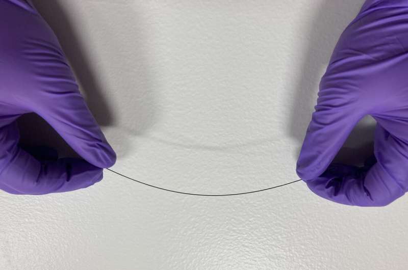
Researchers have created a flexible needle-like endoscopic imaging probe that can acquire 3D microscopic images of tissue. The bendability is possible thanks to a new flexible graded index (GRIN) lens developed by the researchers.
GRIN lenses, originally developed for the telecommunications industry, are commonly used to perform fluorescence microscopy approaches that can image deep into tissues. However, the fact that they are rigid components has limited their use clinically.
"When a traditional biopsy is performed, it represents a single moment in time and can take days to get results back from the laboratory," said research lead Guigen Liu from Harvard Medical School. "Our bendable imaging probes could shorten the waiting time to minutes and enable new approaches that use imaging to dynamically monitor tissue changes, for instance, how tumors react to treatments over time."
In Optics Express, the researchers describe their new GRIN lens, which they incorporated into an endoscopy probe. Experiments showed that the probe's imaging properties are maintained even when it is bent.
"The bendable nature of these GRIN probes makes measurements in living subjects, such as animals or human patients, much more streamlined and practical," said Liu. "It could be useful for precise, minimally invasive microscopy-guided placement of needles and catheters for tissue biopsies and tumor ablation, for example."
Imaging through a bent lens
GRIN lenses are silica glass rods with a continuously changing refractive index that focuses light coming through the rod without requiring a separate focusing lens. Since their development about 50 years ago, it has been generally thought that GRIN lenses can only be used as rigid imaging probes. In the new work, the researchers decided to challenge this notion by finding out if it was possible to image through a bent GRIN lens.
The researchers custom-designed a GRIN lens 500 microns in diameter and about 100 mm long. The lens' long, thin shape and its lack of a rigid outer casing gives it the flexibility to bend about 10 degrees without breaking. They then incorporated the new GRIN lens into an endoscopic imaging probe and tested it by performing two-photon 3D fluorescence imaging through it.
To simulate the real-world bending that would be experienced deep inside tissues, the lens was positioned vertically and pushed to introduce the type of beam deflection that would be experienced if the probe was used in the working channel of a needle used for a biopsy.
The experiment showed that the resolution and signal level did not obviously deteriorate when one end of the probe was displaced laterally by 6 mm. "When the lens is bent, the signal lanes that carry the image through the rod synergistically adapt by laterally shifting, much like a car tends to shift toward the outside of a slippery curved road," said Liu. "While the signal lanes distort a bit while shifting, they largely keep their properties such as order and shape. This allows most of the resolution and signal level to be retained."

A new way to screen cancer drugs
Although further development and testing would be needed to bring the endoscope into the clinic, the device is already finding applications in biomedical research. Team lead and co-author Oliver Jonas and colleagues are pairing the endoscope with a new type of microdevice to test a method for quickly evaluating the effectiveness of various cancer therapies.
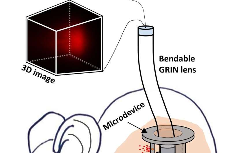
The team's new microdevices are designed to be implanted directly into a tumor and carry small amounts of up to 20 drugs. To measure the effectiveness of the various drugs without removing any tumor tissue, the researchers insert a GRIN-based endoscope directly into the microdevice where it can be used to image fluorescence signals inside the tumor. Although this setup is currently being studied in mice, it could eventually be used in patients to quickly figure out which treatment options are best for fighting each patient's specific tumor.
To move the probes toward clinical application, the researchers are also working to develop longer bendable GRIN lenses to allow deeper imaging and more flexibility. They also want to enhance the mechanical durability of the optical components using a thin polymer coating that won't affect flexibility.
More information: Guigen Liu et al, Bendable long graded index lens microendoscopy, Optics Express (2022). DOI: 10.1364/OE.468827
Journal information: Optics Express
Provided by Optica


