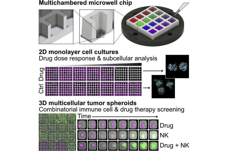
Graphical abstract. Credit: Cell Reports Methods (2022). DOI: 10.1016/j.crmeth.2022.100256
In a study recently published in Cell Reports Methods, co- authors Björn Önfelt, Niklas Sandström and Valentina Carannante, researchers at SciLifeLab and the Department of Microbiology, Tumor and Cell Biology at Karolinska Institutet, describe a new miniaturized method for high-content screening combined with high-resolution imaging, all in the same microchip.
The platform was used to perform drug screening of conventional 2D cell cultures and 3D tumor spheroids. It also tested natural killer (NK) cell responses against tumor spheroids, and how that could be boosted by additional treatment, for example by monoclonal antibodies against tumor antigen and chemotherapeutic drugs. The platform could be used as a complementary tool to test therapeutic strategies, tailoring the treatments to the patient need and to study the mechanisms of tumor progression and drug response.
"The study shows mainly proof-of-principle experiments, but we are currently following this up with studies where the method is used to study donor-dependence in NK cell response to checkpoint blockade and NK cell responses to primary sarcoma," says Björn Önfelt.
"We are already using the method in other projects to show that it could be of clinical or even commercial value, and if we reach clinical implementation or use the platform for drug discovery and development, it may have an immediate impact on treatment and human health," he adds.
More information: Niklas Sandström et al, Miniaturized and multiplexed high-content screening of drug and immune sensitivity in a multichambered microwell chip, Cell Reports Methods (2022). DOI: 10.1016/j.crmeth.2022.100256
Journal information: Cell Reports Methods
Provided by Karolinska Institutet


