New nanotech imaging tool may allow smartphone disease diagnosis

Scientists have developed a low-cost microscopic imaging device small enough to fit on a smartphone camera lens, with the potential to make mobile medical diagnosis of diseases affordable and accessible.
Research published today in ACS Photonics from researchers at the University of Melbourne and the Australian Research Council Centre of Excellence for Transformative Meta-Optical Systems (TMOS), is helping miniaturize phase-imaging technology using metasurfaces, which are only a few hundred nanometers thick—about 350 times thinner than the thickness of a human hair—thus small enough to fit in the lens of a smartphone or other small camera.
The detection of diseases often relies on optical microscope technology to investigate changes in biological cells. Currently, these investigation methods usually involve staining the cells with chemicals in a laboratory environment as well as using specialized "phase-imaging" microscopes. These aim to make invisible aspects of a biological cell visible, so early-stage detection of disease becomes possible. However, phase-imaging microscopes are bulky and cost thousands of dollars, putting them out of reach of remote medical practices.
In addition to providing resources for remote medical practices, this new technology could one day lead to at-home disease detection, where the patient could obtain their own specimen through saliva or a pinprick of blood, and then transmit an image to a laboratory anywhere in the world. The lab could then analyze and diagnose the illness.
Lead researcher, University of Melbourne Dr. Lukas Wesemann said similar to expensive phase-imaging microscopes, these metasurfaces can manipulate the light passing through them to make otherwise invisible aspects of objects like live biological cells visible.
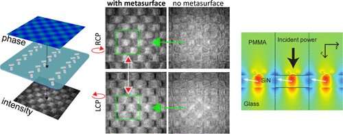
"We manufactured our metasurface with an array of tiny nanorods on a flat surface, arranged in such a way as to turn an invisible property of light, called its 'phase,' into a normal image visible to the human eye, or conventional cameras," Dr. Wesemann said.
"These phase-imaging metasurfaces create high contrast, pseudo-3D images without the need for computer post-processing.
"Making medical diagnostic devices smaller, cheaper and more portable will help disadvantaged regions gain access to healthcare that is currently only available to first world countries."
Co-author, TMOS Chief Investigator and University of Melbourne Professor Ann Roberts, said it was an exciting breakthrough in the field of phase-imaging.
"It's just the tip of the iceberg in terms of how metasurfaces will completely reimagine conventional optics and lead to a new generation of miniaturized devices."
More information: Lukas Wesemann et al, Real-Time Phase Imaging with an Asymmetric Transfer Function Metasurface, ACS Photonics (2022). DOI: 10.1021/acsphotonics.2c00346
Journal information: ACS Photonics
Provided by University of Melbourne


