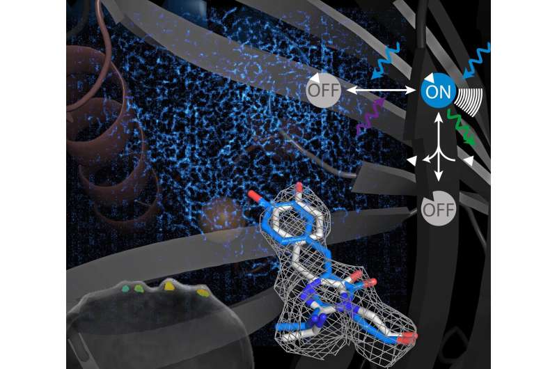
Model of a switchable sensor. Credit: Helmholtz Munich / Andre C. Stiel
Dr. Andre C. Stiel leads the group for Cell Engineering at the Institute of Biological and Medical Imaging at Helmholtz Munich. He contributed to the paper "Genetically encoded photo-switchable molecular sensors for optoacoustic and super-resolution imaging" published in Nature Biotechnology, and explained its relevency in a recent interview:
How do proteins help in biomedical imaging?
Stiel: Proteins are molecules made out of amino acids. In our bodies and in all life forms, proteins have many functions—for example they are the machinery that allows the cells to generate energy or receive information. In imaging, we can utilize proteins that generate signals, for example light, to observe what is happening within a live organism. Proteins have the advantage that they are genetically encoded, they can become a component of the cell or the organism—in contrast to, for example, a dye that needs to be added from the outside. In imaging we can use this for something we call labeling. Labels open up exciting possibilities for tracking cells in different states over a long period of time and without having to interfere or damage the living tissue. This allows us, for example, to study a disease or treatment in a relatively natural way and thus to understand diseases better.
What's the secret of the switchable proteins that you engineer?
Stiel: Switchable label proteins can change their state, in our case the signal that we read out, upon illumination with different colors of light. The molecular mechanism acts like a tiny switch that changes the state of the protein from on to off and with that the signal it generates. In nature, those proteins are often responsible for light dependent responses—for example of plants orienting towards the light.
You use switchable proteins in a method called optoacoustic imaging—what is that?
Stiel: Optoacoustic imaging—where world-leading research is done at Helmholtz Munich by Vasilis Ntziachristos—is an imaging method that relies on reading out ultrasound signal generated by light. Optoacoustic imaging already has the power to deliver a combination of higher penetration depth, a higher resolution and larger fields of view than other imaging technologies. However, for many research questions optoacoustics needs tools like genetically encoded reporters and sensors. Photoswitchable label proteins can help here. In our research, the light switchable signal enables us to visualize small numbers of cells against a strong background of other signals by making the label blink. You can imagine it as a lighthouse in a stormy dark night at sea. The ability to visualize few cells in a live organism is important because many biological phenomena, especially in the immune system, rely on a small number of cells. Our aim is to one day track single labeled cells in a living organism and visualize their function—to improve our knowledge, for example, about the immune system or about tumor development.
What's your next challenge?
Stiel: We talked about visualizing cells. However, cells themselves host even smaller components of life: small molecules and ions. Often such molecules fulfill very dedicated purposes like communication, or serving as nutrient sources or building blocks for other cellular components, hence they need to be tightly regulated for the cell to function properly. Understanding this regulation is essential to understand life and diseases. In order to visualize small molecules or ions, we don't use labels but sensors. Sensors can be envisioned like labels that are only visible if the molecule of interest is present—thus, this allows us to visualize the molecules within a cell. In our latest study, we applied our photoswitching concept to sensors. We showed that photoswitching sensors work for optoacoustic as well as for super resolution. Indeed, the application of switchable proteins is not limited to optoacoustics but they are also important tools in super resolution fluorescence microscopy. We developed a concept and a first prototype—with potential for further optimization. Together with Dierk Niessing from Helmholtz Munich we learned the details of the molecular mechanism and together with a group from KTH in Sweden (Ilaria Testa) we made the first super-resolution images using this concept. The sensors will eventually allow us to also visualize small molecule or ion distributions at nanometer resolution. For optoacoustics our goal is to use them to follow small molecules in a whole live animal—that's the challenge for the next five years.
More information: Kanuj Mishra et al, Genetically encoded photo-switchable molecular sensors for optoacoustic and super-resolution imaging, Nature Biotechnology (2021). DOI: 10.1038/s41587-021-01100-5
Provided by Helmholtz Association of German Research Centres


