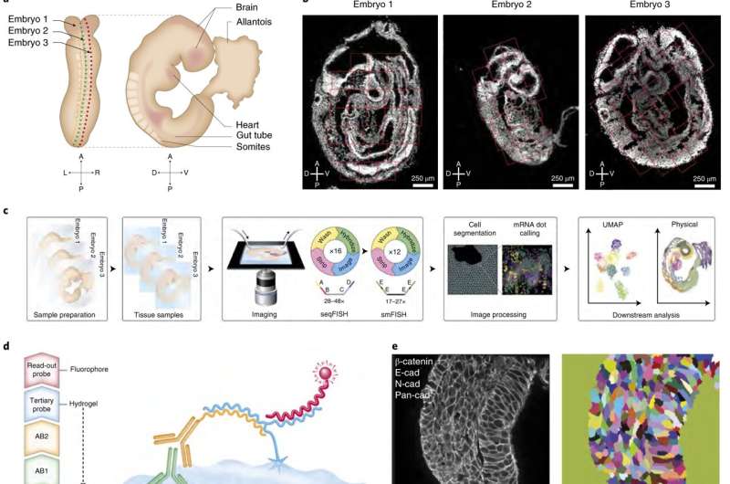
Fig. 1: Single-cell spatial transcriptomics map of mouse organogenesis using seqFISH. a, Illustration of 8–12 ss mouse embryo. Dotted lines indicate the estimated position of the sagittal tissue section shown in b; D, dorsal; V, ventral; R, right; L, left; A, anterior; P, posterior. b, Tile scan of a 20-µm sagittal section of three independently sampled 8–12 ss embryos stained with nuclear dye DAPI (white). Red boxes indicate the selected field of view (FOV) imaged using seqFISH. c, Illustration of the experimental overview for spatial transcriptomics using seqFISH for 351 selected genes in 16 sequential rounds of hybridization and 12 non-barcoded sequential smFISH hybridization rounds for 36 genes. For each targeted gene, 17–48 unique probes were used to capture the mRNA; UMAP, uniform manifold approximation and projection. d, Cell segmentation strategy using a combination of E-cadherin (E-cad), N-cadherin (N-cad), pan-cadherin (Pan-cad) and β-catenin antibody (AB; green) staining detected by an oligo-conjugated anti-mouse IgG secondary antibody (orange) that gets recognized by a tertiary probe sequence. The acrydite group (blue star) of the tertiary probe (blue) gets crosslinked into a hydrogel scaffold and stays in place even after protein removal during tissue clearing. The cell segmentation labeling can be read by a fluorophore-conjugated readout probe (red); AB1, antibody 1; AB2, antibody 2. e, Cell segmentation staining of a 10-µm thick transverse section of an E8.5 mouse embryo using the strategy introduced in d. Cell segmentation signal was used to generate a cell segmentation mask using Ilastik (right). This was repeated independently for all N = 3 embryos with similar results. f, Representative visualization of normalized log expression counts of 12 selected genes measured by seqFISH to validate performance. This experiment was repeated independently for all N = 3 embryos with similar results. g, Highly resolved ‘digital in situ’ of the cardiomyocyte marker titin (Ttn), Tbx5, Cdh5 and Dlk1, colored in red, cyan, green and orange, respectively. Dots represent individually detected mRNA spots, and the box represents an area that was magnified for better visualization. This experiment was repeated independently for all N = 3 embryos with similar results. Credit: DOI: 10.1038/s41587-021-01006-2
High-resolution gene expression maps have been combined with single-cell genomics data to create a new resource for studying how cells adopt different identities during mammalian development. The Spatial Mouse Atlas is the result of a collaboration between researchers at EMBL's European Bioinformatics Institute (EMBL-EBI), the Babraham Institute, the Cambridge Stem Cell Institute, the Cancer Research UK (CRUK) Cambridge Institute, and the California Institute of Technology (Caltech) and colleagues.
Cell fate decisions determine how cells develop into different cell types. The development of different cell types eventually leads to the formation of all the different tissues in the body. This complex process involves many different signals from surrounding tissues, as well as mechanical constraints, epigenetic modifications and changes to gene expression. These factors create unique cell and tissue types, which eventually give rise to all major organs in a process called organogenesis.
Studies using single-cell RNA sequencing (scRNAseq) have provided insights into how the molecular landscape of cells in the mouse embryo changes during early development. However, scRNAseq methods require the cells in the embryo to be dissociated—or separated—which means spatial information is lost.
This study, published in Nature Biotechnology, combines scRNAseq data with spatially-resolved expression profiles to generate an atlas of gene expression at single-cell resolution across the entire embryo.
Combining computational and image-based data
"Methodologically, I think this is one of the most exciting examples of integrating spatially resolved transcriptomics and single-cell sequencing to create a new resource for the scientific community," said John Marioni, Head of Research at EMBL-EBI. "It's also work we can build upon to include more target genes across different stages of development."
Cells organised according to their transcript data changing to the seqFISH mouse embryo map. Credit: Shila Ghazanfar, Cancer Research UK Cambridge Institute. Design: Karen Arnott, EMBL-EBI
The researchers carried out this study using 8–12 somite stage mouse embryos. This stage of development was of particular interest because it is when the cells within the embryo start to differentiate, or become a specific cell type. The researchers applied an image-based single-cell transcriptomics method—seqFISH (developed by collaborators at Caltech) – to detect 387 target genes. They combined this information with scRNAseq data to produce an atlas of cell types found across this stage of mouse development.
"For this study we needed to process, analyze and integrate a lot of data sets to enable us to map the identities of cells onto a spatial reference," says Shila Ghazanfar, Research Associate at CRUK Cambridge Institute. "This new approach means that we can take a section of the embryo and essentially paint on the cell types using different colors to place cell type context on the anatomy of the embryo at single-cell resolution."
Insights into brain and gut development
Tim Lohoff, a Ph.D. student at the Babraham Institute and lead author on the paper, commented: "Previously, researchers have been able to measure gene expression profiles for embryonic cells. However, it has long been a technological challenge to link these profiles to their original spatial location. Our work has been able to overcome this challenge, providing critical information about how the spatial environments of cells are critical to organ development, and opening up new avenues of research."
The researchers were able to use the Spatial Mouse Atlas they created to uncover new insights into mouse development. They were able to find previously unreported gene expression information within the developing brain and gut tube of the mouse. This new approach provides a robust framework for future studies looking at spatial gene expression, both in the mouse and potentially other biological systems.
"This landmark study has already allowed us to pinpoint some of the molecular events that lead to the development of organs in mice. By providing a more detailed blueprint for development shared between mammals, this study could help to further advance the possibility of generating cell types in a dish for regenerative medicine." says Professor Wolf Reik, Group leader in the Epigenetics research program at the Babraham Institute.
More information: T. Lohoff et al, Integration of spatial and single-cell transcriptomic data elucidates mouse organogenesis, Nature Biotechnology (2021). DOI: 10.1038/s41587-021-01006-2
Journal information: Nature Biotechnology
Provided by European Molecular Biology Laboratory


