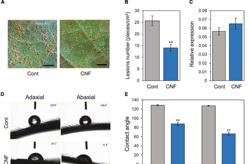
Figure 1. Phakopsora pachyrhizi lesion formation, pre-infection structures formation, and hydrophobicity on CNF-treated soybean leaves. Disease lesions (A) and lesion numbers (B) resulting from P. pachyrhizi infection on the abaxial leaf surface of control, and leaves covered with 0.1% cellulose nanofiber (CNF). Soybean plants were spray-inoculated with P. pachyrhizi (1 × 10⁵ spores/ml). Photographs were taken 10 days after inoculation. Bars indicate 0.2 cm. Lesion numbers were counted to calculate lesion number per cm2. Vertical bars indicate the standard error of the means (n = 54). Asterisks indicate a significant difference between control and CNF-treatments in a t-test (**p < 0.01). (C) Urediniospore attachment quantification on the leaf surface of control and leaves covered with 0.1% CNF derived. Soybean plants were spray-inoculated with P. pachyrhizi (1 × 10⁵ spores/ml) and immediately total RNAs including soybean and P. pachyrhizi were purified. Relative expression of soybean ubiquitin 3 (GmUBQ3) and P. pachyrhizi ubiquitin 5 (PpUBQ5) were evaluated using RT-qPCR. Vertical bars indicate the standard error of the means (n = 4). Droplet profiles (D) and quantification of contact angles (E) on the adaxial and abaxial leaf surface of control, and leaves covered with 0.1% CNF derived. Contact angles were evaluated as described in section “Materials and Methods”. Vertical bars indicate the standard error of the means (n = 60). Asterisks indicate a significant difference between control and CNF-treatments in a t-test (**p < 0.01). P. pachyrhizi pre-infection structure formation (F) and percentage of urediniospores (G) on the adaxial and abaxial surfaces of control, and leaves covered with 0.1% CNF, treated with 0.1% DMSO and 500 mM scopoletin (Sco). Soybean plants were spray-inoculated with P. pachyrhizi (1 × 10⁵ spores/ml). The pre-infection structures were stained with Calcofluor White and photographs were taken 6 h after inoculation. Bars indicate 50 μm. The percentage of germinated (Ge) urediniospores and differentiated germ-tubes with appressoria (Ap) were evaluated as described in section “Materials and Methods.” Vertical bars indicate the standard error of the means (n = 21). Significant differences (p < 0.05) are indicated by different letters based on a Tukey’s honestly significant difference (HSD) test. Credit: DOI: 10.3389/fpls.2021.726565
A water-absorbent coat to keep rust away? It may seem counterintuitive but when it comes to soybean plants and rust disease, researchers from Japan have discovered that applying a coating that makes leaf surfaces water absorbent helps to protect against infection.
In a study published this month in Frontiers in Plant Science, researchers from the University of Tsukuba have revealed that coating soybean plant leaves with cellulose nanofiber changes the leaf surface from water repellent to water absorbent and offers resistance against Asian soybean rust.
Rusts are plant diseases that get their name from the powdery rust- or brown-colored fungal spores on the surfaces of infected plants. Asian soybean rust (ASR) is an aggressive disease of soybean plants, causing estimated crop yield losses of up to 90%. ASR is caused by Phakopsora pachyrhizi, a fungal pathogen that requires a living plant host to survive. The timely application of fungicide is currently the only way of controlling ASR in the field. But the use of fungicides can be problematic, resulting in negative environmental effects, increased production costs, and fungicide-resistant pathogens.
"We investigated cellulose nanofiber (CNF) as an alternative method of controlling ASR," says senior author of the study, Professor Yasuhiro Ishiga. "Specifically, we wanted to know whether coating soybean plant leaves with CNF protected plants against P. pachyrhizi."
Of the available methods for isolating CNF, aqueous counter collision (ACC) has been shown to alter the hydrophilic (water absorbent) and hydrophobic (water repellent) properties of surfaces, switching one to the other. Previous research has indicated that CNF obtained via ACC has higher wettability than CNF isolated by other methods.
"We showed that CNF can change the soybean leaf surface from hydrophobic to hydrophilic," explains senior author, Professor Yuji Yamashita. "This offers resistance against P. pachyrhizi."
The team found fewer lesions and significantly reduced formation of P. pachyrhizi appressoria, which are specialized pre-infection structures used to break through the outer surface of the host plant, on CNF-treated leaves compared with control (untreated) leaves. The results also revealed suppressed gene expression linked to the formation of pre-infection structures in P. pachyrhizi on treated versus control leaves.
"In particular, chitin synthase gene expression was suppressed, and P. pachyrhizi needs chitin synthases to form pre-infection structures," says Professor Ishiga.
This study is the first to investigate the application of CNF for controlling plant diseases in the field, and this technique offers new possibilities for sustainable and eco-friendly management of plant diseases.
More information: Haruka Saito et al, Covering Soybean Leaves With Cellulose Nanofiber Changes Leaf Surface Hydrophobicity and Confers Resistance Against Phakopsora pachyrhizi, Frontiers in Plant Science (2021). DOI: 10.3389/fpls.2021.726565
Journal information: Frontiers in Plant Science
Provided by University of Tsukuba


