Improving the observation of protein bonding

Researchers working with Oak Ridge National Laboratory developed a new method to observe how proteins, at the single-molecule level, bind with other molecules and more accurately pinpoint certain molecular behavior in complex physiological environments.
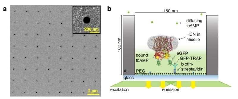
At ORNL's Center for Nanophase Materials Sciences, researchers produced zero-mode waveguides, or ZMWs, which are optical tools composed of aluminum films etched with small holes used to illuminate selected areas of a sample and eliminate background noise.
"Because the aperture is so small, the field only extends in that pocket or just above that pocket," said ORNL's Scott Retterer. "You're only getting signal from that extremely small area within the aperture and everything else is dark."
Using ORNL-produced ZMWs, researchers from the Washington University in St. Louis, School of Medicine and the University of Wisconsin-Madison used the method to observe proteins that are implicated in human heart function.
More information: David S. White et al, cAMP binding to closed pacemaker ion channels is non-cooperative, Nature (2021). DOI: 10.1038/s41586-021-03686-x
Journal information: Nature
Provided by Oak Ridge National Laboratory


