A new polarized fluorescent probe for revealing architectural dynamics of living cells

Monitoring alignments of the building blocks of cells is important to understand how the cells are built. By collaborating with imaging scientists at the MBL, researchers from Japan have developed a new probe which they call POLArIS, allowing real-time imaging of molecular orientations in live cells.
A fluorophore emits polarized light as it glows. The orientation of polarized fluorescence is closely related to the orientation of the fluorophore. If a molecule of interest is rigidly connected with a fluorescent tag such as Green Fluorescent Protein (GFP), the polarized fluorescence from the fluorophore reports the orientation of the molecule.
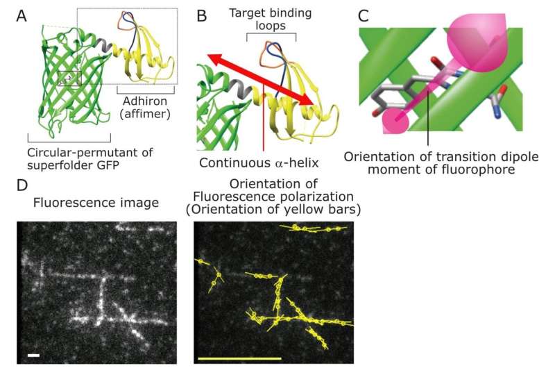
"In previous approaches for monitoring the orientation of the protein of interest, researchers needed to develop effective constrained GFP tagging methods which might be different for each protein of interest," says one of the lead authors of the study, Ayana Sugizaki. "POLArIS uses an antibody-like binder that is rigidly connected with GFP, allowing both specific and versatile constrained labeling," adds another lead author Keisuke Sato. The team used a commercially available Adhiron molecule (now rebranded as "Affimer") as the binder molecule to link GFP to a target protein, and developed POLArIS by connecting Adhiron and GFP in a rotationally constrained manner (Figure 1).
Because an Adhiron molecule that specifically binds to a molecule of interest can be easily selected from a library of molecules through phage display screening, POLArIS can be designed for any biological molecules of interest. POLArIS can be expressed in specific cell types and organelles, and will be useful for studying architectural dynamics of molecular assemblies in broad range of cell cultures, tissues and whole organisms. "From the point of view of fluorescence polarization imaging, POLArIS has significant advantages because of its genetically encoded nature," says Tomomi Tani, a Senior Researcher at the National Institute of Advanced Industrial Science and Technology (AIST) in Japan, who has joined this project since he was an Associate Scientist at the MBL.
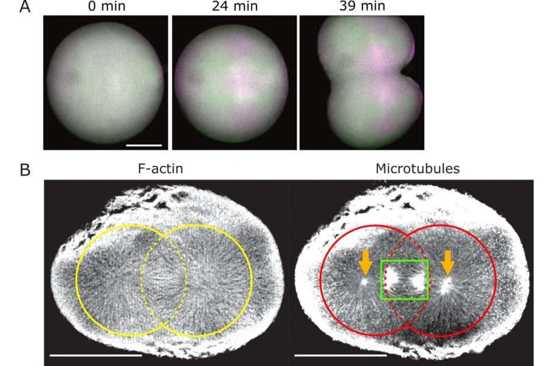
By using the probe for actin, the team uncovered transient emergence and dissolution of highly ordered F-actin architecture that they named FLARE structure, in dividing cells of starfish embryo (Figure 2 and Movie 1). "We found that the structure extends up to the cell cortex in association with the astral microtubules," says corresponding author, Sumio Terada, who had frequently visited the MBL from Tokyo, together with his colleagues in TMDU. "The astral microtubules are responsible for connecting the spindle to the cell cortex and orientating it correctly, controlling the plane of cell divisions." The mechanisms that determine the cell division plane is the key for controlling many aspects of development, and yet a crucial mystery. The discovery of radially aligned actin architectures will shed light on the most fundamental unanswered questions of cell biology.

More information: Ayana Sugizaki et al, POLArIS, a versatile probe for molecular orientation, revealed actin filaments associated with microtubule asters in early embryos, Proceedings of the National Academy of Sciences (2021). DOI: 10.1073/pnas.2019071118
Journal information: Proceedings of the National Academy of Sciences
Provided by Tokyo Medical and Dental University


