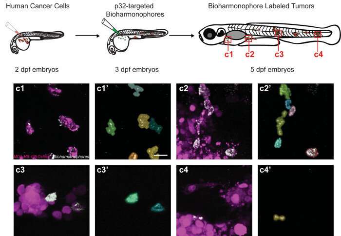
New Imperial-developed nanoprobes tested in zebrafish could help detect cancer more accurately and might aid diagnosis and therapy in the future. Credit: Imperial College London
To highlight tumors in the body for cancer diagnosis, doctors can use tiny optical probes (nanoprobes) that light up when they attach to tumors. These nanoprobes allow doctors to detect the location, shape and size of cancers in the body.
Most nanoprobes are fluorescent; they absorb light of a specific color, like blue and then emit back light of a different color, like green. However, as tissues of the human body can emit light as well, distinguishing the nanoprobe light from the background light can be tough and could lead to the wrong interpretation.
Now, researchers at Imperial College London have developed new nanoprobes, named bioharmonophores and patented at Imperial, which emit light with a new type of glowing technology known as second harmonic generation (SHG).
After testing the nanoprobes in zebrafish embryos, the researchers found that bioharmonophores, which were modified to target cancer cells, highlighted tumors more brightly and for longer periods than fluorescent nanoprobes. Their light can be easily spotted and distinguished by the tissue generally emitted light, and they also attach precisely to tumor cells and no healthy cells, making them more precise in detecting tumor edges.
Lead researcher Dr. Periklis Pantazis of Imperial's Department of Bioengineering said: "Bioharmonophores could be a more effective way to detect tumors than is currently available. They uniquely combine features that could be great for cancer diagnosis and therapy in clinical practice and could eventually improve patient outcomes following further research."
The findings are published in ACS Nano.
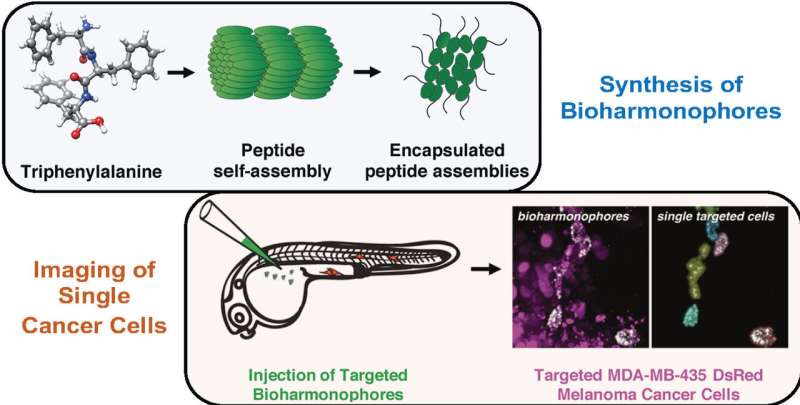
Bioharmonophores are both biocompatible and biodegradable as they are made of peptides—the same ingredients of proteins found in the body. They are metabolized naturally in the body within 48 hours and are therefore unlikely to pose long-term health risks.
Bioharmonphores are relatively simple to assemble. Credit: Imperial College London
To investigate precise tumor detection, the researchers first injected zebrafish embryos with malignant cancer cells, which allowed tumor cells to proliferate unchecked. Twenty-four hours later they injected bioharmonophores which were modified to target p32 peptide molecules that are specifically found in tumor cells. They then used imaging techniques at Imperial's Facility for Imaging by Light Microscopy to study how well the modified bioharmonophores detected the tumors.
They found that bioharmonophores had outstanding detection sensitivity, meaning they attached to specific tumor cells but not to healthy ones. Fluorescence-enabled nanoprobes tend to attach less specifically, meaning they can misrepresent healthy cells as tumor cells, or vice versa.
They also found that unlike fluorescence, bioharmonophores did not 'bleach', meaning they did not lose their ability to emit light over time. In addition, the light emitted by bioharmonophores did not saturate as happens with fluorescent nanoprobes, meaning they got brighter when illuminated with more light. This way tumors became even clearer.
Dr. Pantazis said: "It is very important that tumor nanoprobes highlight cells specifically and clearly for cancer diagnosis. Our proof-of-concept study suggests that the very bright bioharmonophores could be powerful tools in diagnosing cancer and targeting treatments in the coming years."
The manufacture of bioharmonophores is cheap, reproducible, scalable and takes around two days at room temperature. They now need to be tested in mammals to identify how well the findings translate beyond zebrafish.
The researchers are also looking into how bioharmonophores could be used to guide surgical interventions during cancer surgery, as well as how they could generate light at different frequencies to potentially help kill tumor cells with high precision.
"Biodegradable Harmonophores for Targeted High-Resolution In Vivo Tumor Imaging" by Ali Yasin Sonay, Konstantinos Kalyviotis, Sine Yaganoglu, Aysen Unsal, Martina Konantz, Claire Teulon, Ingo Lieberwirth, Sandro Sieber, Shuai Jiang, Shahed Behzadi, Daniel Crespy, Katharina Landfester, Sylvie Roke, Claudia Lengerke, and Periklis Pantazis, published 25 February 2021 in ACS Nano.
More information: Ali Yasin Sonay et al. Biodegradable Harmonophores for Targeted High-Resolution In Vivo Tumor Imaging, ACS Nano (2021). DOI: 10.1021/acsnano.0c10634
Journal information: ACS Nano
Provided by Imperial College London


