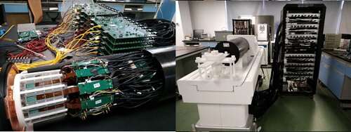
Photo of SIAT aPET. Credit: SIAT
Positron emission tomography (PET) is an important tool for studying the animal model of human diseases and the development of new drugs and new therapies.
The spatial resolution and sensitivity are two important parameters that affect the quantitative accuracy of PET studies.
A research team led by Prof. Yang Yongfeng, and the co-first authors Kuang Zhonghua and Dr. Wang Xiaohui from the Shenzhen Institutes of Advanced Technology (SIAT) of the Chinese Academy of Sciences developed a small animal PET scanner named SIAT aPET with high spatial resolution and high sensitivity.
The study was published in Physics in Medicine & Biology.
Due to the uncertainty of the depth of interaction of traditional PET detectors, small animal PET scanner cannot achieve a high spatial resolution and a high sensitivity simultaneously.
In this work, the researchers developed and used high resolution depth encoding PET detectors using dual-ended readout of pixeled LYSO array with SiPM arrays. The scanner achieved a sensitivity of 16% at the center of field of view and an average spatial resolution of 0.82 mm in a field of view large enough for imaging the rats, both of which are outstanding results.
"The scanner has been very stable. A few animal studies were already performed on the scanner and we are expecting to do more animal scans very soon," said Prof. Yang. "The scanner is also designed to be MRI compatible and recent MRI-PET interference test experiments showed very promising results. We believe the scanner will improve the quantitative accuracy of the PET studies and reduce the scan time."
More information: Zhonghua Kuang et al. Design and performance of SIAT aPET: a uniform high-resolution small animal PET scanner using dual-ended readout detectors, Physics in Medicine & Biology (2020). DOI: 10.1088/1361-6560/abbc83
Journal information: Physics in Medicine and Biology
Provided by Chinese Academy of Sciences


