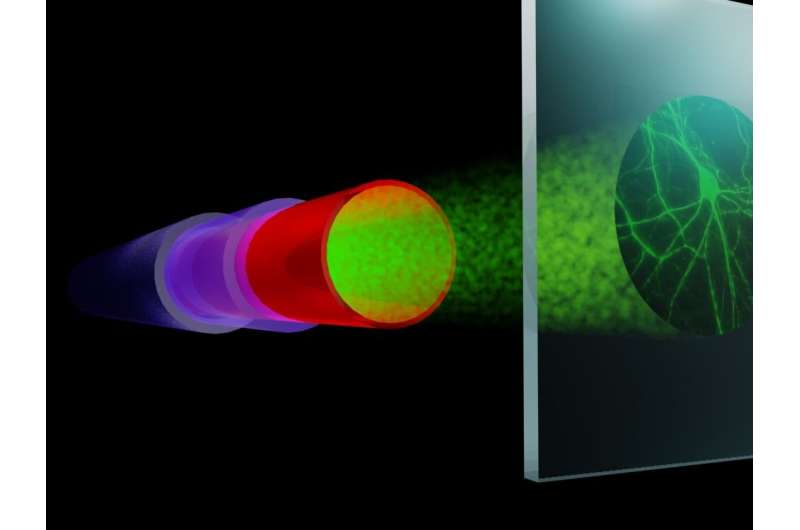
Artist impression of the super-resolution fiber setup. A randomly speckled beam (green) from the fiber illuminates the entire sample (right) several times. Compressive sensing reconstruction offers a high-resolution image of the sample without the need for fluorescent labeling, offering nanoscopy applications in both bioimaging and nanolithography. Credits: Lyuba Amitonova
Researchers at ARCNL and Vrije Universiteit Amsterdam have developed a compact setup for fast, super-resolution microscopy through an ultrathin fiber. Using smart signal processing, they beat the theoretical limits of resolution and speed. Because the method does not require any special fluorescent labelling, it is promising for both medical applications and characterization of 3-D structures in nanolithography. On May 7th, the results were published in Light: Science & Applications, a scientific journal in the Nature family.
"Imaging at the nanoscale is limited by the wavelength of the light that is used. There are ways to overcome this diffraction limit, but they typically require large microscopes and difficult processing procedures," says Lyuba Amitonova. "These systems are unsuitable for imaging in deep layers of biological tissue or in other hard-to-reach places."
Amitonova recently started a research group on Nanoscale Imaging and Metrology at ARCNL. She is also connected part-time to VU Amsterdam where she works on ultrathin fibers for endomicroscopy in the group of Johannes de Boer. Amitonova and de Boer have developed a way to overcome the diffraction limit in small systems to enable deep-tissue imaging with super-resolution.
Inverse data compression
Key to Amitonova's approach is the fact that not all of the information in a data sample is needed to create a meaningful image. "Think of digital photography, which uses the JPEG compression format to limit the amount of data in a picture. The compression removes up to ninety percent of the image, but we can hardly see the difference," she says. "This works, because all conventional images of real-life objects are 'sparse,' which means that most image points do not contain any information. In our measurements, we use this sparsity of information in an inverse way, by acquiring only ten percent of the available data and reconstructing the entire image via a mathematical computation method."
Speckled beam
In conventional microscopy, samples are often illuminated point by point to create a picture of the entire sample. This takes a lot of time, as high-resolution images require many data points. The approach developed by Amitonova and de Boer uses a fiber that produces a speckled laser beam, which allows to illuminate many areas in the sample simultaneously in a random way. The multifaceted light reflected by the sample is then collected as a single data point, from which relevant information is extracted by computation. "With point by point illumination, taking 256 data points would result in a 256 pixels image. With our method, the same number of measurements creates an image of about twenty times as many pixels," says Amitonova. "Thus, compressive imaging is much faster, but we also demonstrate that it is capable of resolving details that are more than two times smaller than can be resolved by conventional diffraction limited imaging."
Label-free sensing
The method was developed with minimally invasive bioimaging in mind. But it is also very promising for sensing applications in nanolithography, because it does not require fluorescent labeling, which is necessary in other methods of super-resolution imaging. Amitonova will develop the concept further at ARCNL: "The compactness of the fibers makes them very convenient for developing metrology tools in nanolithography. The fiber-based probes provide a unique combination of high resolution with a large field of view and could be easily used in hard-to-reach places. Developing our methods further will hopefully result in even higher resolution and speed. Metrology tools and medical diagnostics are the most likely areas to benefit from our findings."
More information: Lyubov V. Amitonova et al. Endo-microscopy beyond the Abbe and Nyquist limits, Light: Science & Applications (2020). DOI: 10.1038/s41377-020-0308-x
Journal information: Light: Science & Applications , Nature
Provided by Advanced Research Center for Nanolithography


