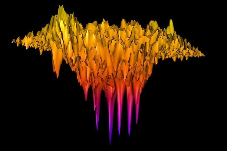
Three-dimensional map of the forces applied by the invading structures of an individual cancer cell. The height of each of the ‘teeth’ seen in the image illustrates the force applied by an invadopod, a small structure underneath the cell that releases enzymes to promote cancer invasion. Credit: University of St Andrews
International research led by the University of St Andrews reveals the first direct measurement of the force cancer cells can apply when they invade, giving insights that may lead to possible future cancer treatments.
The research, in collaboration with the Albert Einstein College of Medicine in New York and published in the journal Science Advances (Wednesday 11 March), also finds that the force that cancer exerts is linked to its ability to attack and degrade healthy tissue in the human body.
Many cancers form invadopodia, tiny protrusions about a thousandth of a millimeter in size, that promote invasion of healthy tissue by releasing enzymes which digest the surrounding tissue.
Although it has been hypothesised that invadopodia exert mechanical force that is implicated in cancer invasion, direct measurements remained elusive. The research, led by Professor Malte Gather from the School of Physics and Astronomy at the University of St Andrews, used cells that form a model for head and neck cancer and an artificial replica of healthy tissue to study the force that cancer cells apply as they invade. Head and neck squamous carcinoma is a cancer found in the mucous membranes of the mouth, nose and throat.
The international team of scientists utilized a recently developed interferometric force imaging technique that provides pico-Newton resolution to quantify invadopodial forces in cells and monitor their temporal dynamics.
The research team compared the force exerted by individual protrusions to their ability to degrade extra cellular matrix, a main component of healthy tissue. They also investigated the mechanical effects of inhibiting invadopodia through genetic modification of the cancer cells.
Professor Malte Gather said: "It has been known for a long time that cancer cells have little spots on their surface that secrete chemicals to degrade the surrounding tissue. Our work now confirms that these spots also push against the tissue, and so most likely act as a chemically assisted mechanical drill allowing cancer to invade healthy cells."
Professors Segall and Prystowsky, from Albert Einstein College of Medicine in New York, said: "The force imaging technology developed at St Andrews combined with cell biologic approaches gives us a new window into ways to elucidate mechanisms of cancer invasion and new possibilities to inhibit tumor cell invasion."
By connecting the biophysical and biochemical characteristics of invadopodia, the study provides a new perspective on cancer invasion which in the future may help to identify biomechanical targets for cancer therapy.
More information: E. Dalaka et al. Direct measurement of vertical forces shows correlation between mechanical activity and proteolytic ability of invadopodia, Science Advances (2020). DOI: 10.1126/sciadv.aax6912
Journal information: Science Advances
Provided by University of St Andrews


