Microscopic spines connect worm neurons

Dendritic "spines"—small protrusions on the receiving side of the connection (synapse) between two nerve cells—are recognized as key functional components of neuronal circuits in mammals. The shapes and numbers of spines are regulated by neuronal activity and correlate with learning and memory.
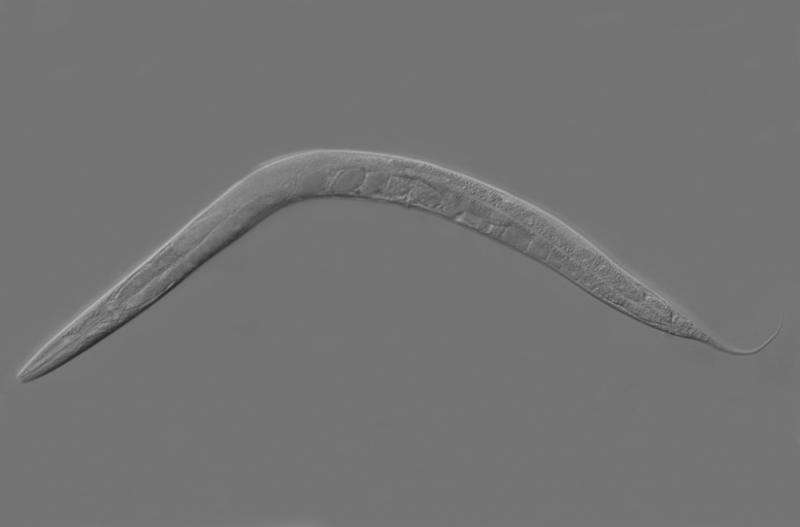
Although spine-like protrusions have been reported in the nervous system of the invertebrate worm C. elegans, it is not known if these structures share functional features with vertebrate dendritic spines.
Now, Andrea Cuentas-Condori, Sierra Palumbos, David Miller, Ph.D., and colleagues have used super-resolution microscopy, electron microscopy, live-cell imaging and genetics to characterize spine-like structures on C. elegans motor neurons. They report in the journal eLife that C. elegans spines are dynamic structures that sense and respond to neuronal activity, like their mammalian counterparts.
The studies establish the genetically tractable and transparent C. elegans as a model organism for the study of dendritic spine formation and function. Live-cell imaging studies and unbiased genetic screens should speed the discovery of genes that regulate spine biology.
More information: Andrea Cuentas-Condori et al. C. elegans neurons have functional dendritic spines, eLife (2019). DOI: 10.7554/eLife.47918
Journal information: eLife
Provided by Vanderbilt University


