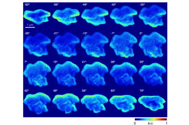
Experimental carbon channel EDS tilt series. All 20 processed carbon EDS projections used as input for GENFIRE reconstruction. Each image was masked, normalized to reference projection, background subtracted, and aligned by center-of-mass and common line alignment. Background subtraction was iterated with alignment to minimize common line differences. Horizontal direction is the rotation axis. Credit: Science Advances, doi: 10.1126/sciadv.aax3009
Multimodal microscopy can combine complementary nanoscale imaging techniques to extract comprehensive information on the chemical, structural and functional aspects of heterogenous samples. X-ray microscopy can achieve high-resolution imaging of bulk materials with chemical, magnetic, electronic and bond orientation contrast. In parallel, electron microscopy can provide spatial resolution at the atomic scale while quantifying the elemental composition.
In a new report, Yuan Huang Lo and colleagues in the interdisciplinary departments of physics, bioengineering and the Advanced Light Source in the U.S. combined X-ray ptychography and scanning transmission X-ray spectromicroscopy (STXM). They then combined the setup with three-dimensional (3-D) energy-dispersive spectroscopy and electron tomography to map the structural and chemical composition of an Allende meteorite particle with 15-nm spatial resolution. The scientists used textural and quantitative elemental information to understand the mineral composition and discuss potential processes before and after accretion (formation of larger astronomical bodies influenced by gravitation).
X-ray and electron microscopy can visualize the structure and function within organic and inorganic systems across spatial scales down to the atomic scale. Advances in X-ray ptychography – a powerful coherent diffractive imaging (CDI) method have extended soft X-ray imaging toward 5-nm spatial resolution. Ptychographic X-ray CDI can image extended integrated circuits and biological structures in two-dimensions (2-D) and 3-D. Scanning transmission X-ray spectroscopy combined with X-ray absorption spectroscopy (XAS) can map bulk specimen with 20-nm resolution, to simultaneously extract chemical-specific maps of carbon, nitrogen, oxygen and transition metals (iron, manganese and nickel). Newly introduced exciting developments in the field, such as cryogenic preservation of biospecimens and the introduction of aberration-corrected electron microscopes have ushered in a new era of cryo-electron microscopy and atomic electron microscopy. These new methods have allowed unprecedented imaging of materials and the associated relationships of structure and function at a fundamental level.
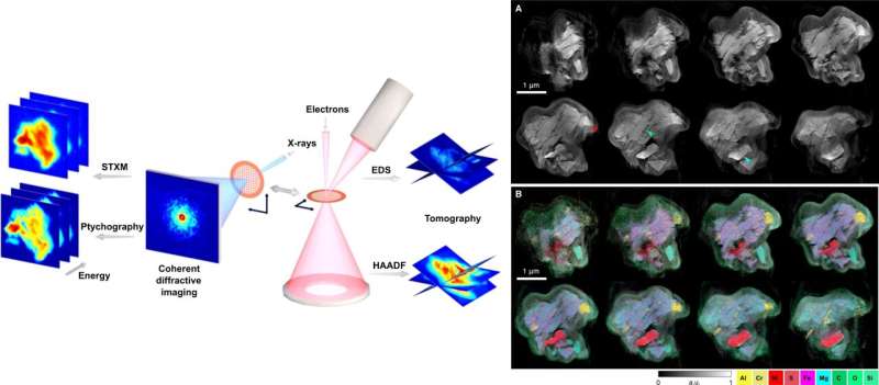
LEFT: Multimodal x-ray and electron nanoscopic spectral imaging scheme. Allende meteorite grains deposited on a TEM grid were transferred between a Titan 60-300 electron microscope and the COSMIC soft x-ray beamline for tomographic, ptychographic, and spectromicroscopic imaging. COSMIC’s TEM-compatible sample holder enabled the same meteorite grain to be imaged using both imaging modalities to extract multidimensional datasets, providing chemical, structural, and functional insights with high spatial resolution. RIGHT: HAADF and EDS GENFIRE tomography reconstructions. Representative 14-nm-thick layers in the reconstructed 3D HAADF (A) and EDS (B) volumes of the Allende meteorite grain. The red arrow points to melt pockets, and the green arrow points to shock veins that were embedded, which suggest that the sample had at some point experienced impact-induced heating, cracking, and melting. The traces of aluminum and chromium in the veins that are visible in the EDS reconstructions reveal that the veins were filled with metallic recrystallization. a.u., arbitrary units. Credit: Science Advances, doi: 10.1126/sciadv.aax3009
Nevertheless, none of the imaging techniques can provide a comprehensive map to simultaneously extract multiple information from a sample. For instance, while electron microscopy can offer unmatched atomic resolution, the method only applies to very thin samples. In comparison, researchers had successfully implemented multimodal imaging in the light microscopy and medical imaging communities. Combining the methods can be effective to prevent sample radiation damage via x-rays, since electrons are more dose efficient than x-rays in elastic scattering experiments. Combined correlative imaging can generate multifaceted experimental maps to guide computational modeling to promote the rapid discovery and deployment of new materials to address challenging scientific questions. The challenges have motivated scientists to incorporate multimodal imaging, with advanced X-ray and electron microscopies to study specimen and take advantage of recent developments in sample imaging.
In the present work, Lo et al. used X-ray ptychography and STXM in 2-D to investigate an Allende meteorite grain. They combined the setup with energy dispersive spectroscopy (EDS) and high-angle annular dark field (HAADF) imaging in 3-D. The Allende meteorite was observed in Mexico on February 8, 1969, as a CV3 carbonaceous chondrite. Researchers studied Allende well at the time as research laboratories were well prepared to receive lunar samples from the Apollo program. They observed the presence of larger chondrules and calcium-aluminum rich inclusions in Allende, set in a fine-grained matrix of micrometer to sub-micrometer-sized silicates, oxides, sulfides and metals. The highly heterogenous meteorite made it an ideal candidate to demonstrate the advantages of multimodal X-ray and electron microscopy.
Lo et al. significantly enhanced the spatial resolution achieved thus far on the meteorite to understand the mineral composition and discuss processes that may have occurred before or after accretion. The imaging results revealed many internal textures and channels to suggest shock veins and melt aggregates on the meteorite. Using the spectroscopic measurements, they classified the major meteoric components as silicates, sulfides and oxides. The multidimensional work provided possible hints to the origins and transport of the Allende meteorite within the early solar nebula and highlighted the potential to combine X-ray and electron imaging to study diverse heterogenous materials.
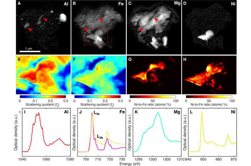
X-ray ptychography and STXM absorption spectromicroscopy. (A to D) Localization of major elements in the meteorite revealed by dividing pre-edge and on-edge ptychography images at the absorption edges for Al, Fe, Mg, and Ni. The absorption quotient maps, displayed in logarithmic scale, show the presence of Fe in the shock veins of the silicate that is barely observable in EDS images (red arrows). (E and F) Scattering quotient (fq) maps derived from ptychographic Mg pre-edge and Al pre-edge images, respectively. This region of interest is a zoomed-in view from the dashed red rectangle shown in (B). (G and H) Ni-Fe ratio maps from Mg pre-edge and Al pre-edge scattering quotient maps, respectively. These ratio maps are converted using the SQUARREL method, given a fixed amount of sulfur. The color bar indicates the Ni-Fe ratio and 100% implies a pure nickel sulfide region. (I to L) Absorption spectra generated from STXM energy scans across the four absorption edges, revealing unique spectral fingerprints for each respective element and also showing pronounced spectral differences in the different iron-containing regions. Relative peak intensities between Fe L3a and L3b also reveal the presence of predominant Fe2+ species. Credit: Science Advances, doi: 10.1126/sciadv.aax3009
The scientists conducted multimodal electron and X-ray spectral imaging using HAADF and EDS tomography and deposited the Allende meteorite grain on a carbon-coated TEM grid and transported it to the COSMIC beamline for X-ray imaging. When they sliced through the generalized Fourier iterative reconstruction (GENFIRE) HAADF of the grain, they observed a variety of internal morphologies that revealed different phases of assembly. In the larger meteorite matrix, they observed long internal channels ranging from 20 to 50 nm in diameter to suggest shock veins. Adjacent to the matrix, they observed two high-intensity spherical granules representing melt pockets. Using EDS tomography, the team determined the elemental composition of the grain and observed the presence of Carbon (C), Oxygen (O), Magnesium (Mg), Aluminum (Al), Silicon (Si), Sulphur (S), Chromium (Cr), Iron (Fe) and Nickel (Ni). They superimposed the EDS and HAADF GENFIRE reconstructions to reveal the main mineral domains, including iron magnesium silicate, aluminum-chromium iron oxide and iron-nickel sulphide.
The research team used differential X-ray absorption contrast to study elemental locations and abundances in more detail to complement the electron microscopy results. For this, they collected 2-D ptychography images of the grain to pinpoint locations of each element with high contrast and spatial resolution. Upon closer inspection, the team observed regions with higher Fe concentrations in the silicate to coincide with the Al melt pockets and veins. The research team developed a SQUARREL (scattering quotient analysis to retrieve the ratio of elements) method to retrieve quantitative information on elemental composition of the complex ptychography images. The scientists obtained two scattering quotient maps for Mg pre-edge and Al pre-edge images as a region of interest. These maps showed new and different image contrast compared to conventional XAS images or phase contrast images. The work distinguished different mineral polymorphs as a strong advantage of the XAS technique compared to conventional EDS and Lo et al. highlighted the complementary nature of X-ray and electron microscopy within the study.
Possible grain composition based on EDS quantification of elemental abundances. Ternary plots of major elements as quantified by the Cliff-Lorimer method for three different mineral types in the meteoric grain. Quantitative compositional information narrows down the possible mineral types and suggests that the sulfide is similar to pentlandite (A), the silicate is similar to ferrosilite (B), and the oxide is a chromium spinel or chromite (C). wt %, weight %. Credit: Science Advances, doi: 10.1126/sciadv.aax3009
In this way, Yuan Huang Lo and a team of researchers combined X-ray and electron microscopy to study the Allende meteorite and yield complementary information on the structural and chemical states of the heterogenous sample. The combined information will assist researchers to identify possible mineral phases present in the meteoric grain with nanometer spatial resolution. The scientists plotted the compositions of major elements in the iron-nickel sulfide, iron-magnesium silicate and aluminum-chromium iron oxide regions of the grain. Using analyses from both X-ray and electron microscopy data, the team narrowed the identities of the various phase groupings to provide a detailed nanoscopic petrographical picture of the meteorite grain.
The team quantified the Ni and Fe composition in the iron-nickel sulfide region using the SQUARREL method (a semiquantitative ptychography analysis method) to reconfirm the identity of the compound, previously only predicted using EDS. The HAADF tomography and X-ray ptychography provided high-resolution textural information on the possible processes that affected the Allende parent body during and after accretion. They explained the possibility of shock melting as the cause for localized shock veins and pockets to generate the highly deformed material. In total, the evidence that Lo et al. gathered using the two imaging techniques (HAADF and EDS with X-ray ptychography and XTSM absorption spectromicroscopy) agreed well with previous 2-D studies of Allende, which were comparatively at a coarser resolution.
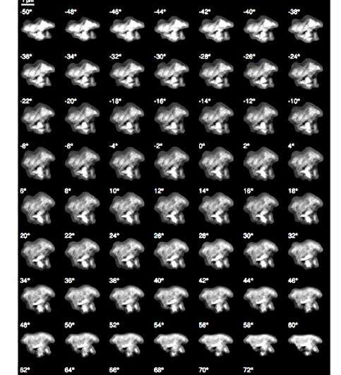
Experimental HAADF tilt series. All 69 processed HAADF projections used as input for GENFIRE reconstruction. Each image was masked, normalized to reference projection, background subtracted, and aligned by center-of-mass and common line alignment. Background subtraction was iterated with alignment to minimize common line differences. Horizontal direction is the rotation axis. Credit: Science Advances, doi: 10.1126/sciadv.aax3009
The researchers highlighted the synergistic relationship between electron and X-ray imaging and their complementary advantages. The multidimensional data gathered in the study provided quantitative chemical and textural information on the diverse phases of the meteorite grain. This multimodal imaging approach is applicable to several other heterogenous systems beyond meteorites to gain new insights across many other interesting and complicated materials.
More information: Yuan Hung Lo et al. Multimodal x-ray and electron microscopy of the Allende meteorite, Science Advances (2019). DOI: 10.1126/sciadv.aax3009
David A. Muller. Structure and bonding at the atomic scale by scanning transmission electron microscopy, Nature Materials (2009). DOI: 10.1038/nmat2380
Franz Pfeiffer. X-ray ptychography, Nature Photonics (2017). DOI: 10.1038/s41566-017-0072-5
Journal information: Science Advances , Nature Materials , Nature Photonics
© 2019 Science X Network


