Team maps magnetic fields of bacterial cells and nano-objects for the first time

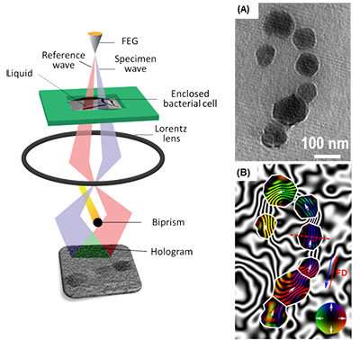
A research team led by a scientist from the U.S. Department of Energy's Ames Laboratory has demonstrated for the first time that the magnetic fields of bacterial cells and magnetic nano-objects in liquid can be studied at high resolution using electron microscopy. This proof-of-principle capability allows first-hand observation of liquid environment phenomena, and has the potential to vastly increase knowledge in a number of scientific fields, including many areas of physics, nanotechnology, biofuels conversion, biomedical engineering, catalysis, batteries and pharmacology.
"It is much like being able to travel to a Jurassic Park and witness dinosaurs walking around, instead of trying to guess how they walked by examining a fossilized skeleton," said Tanya Prozorov, an associate scientist in Ames Laboratory's Division of Materials Sciences and Engineering.
Prozorov works with biological and bioinspired magnetic nanomaterials, and faced what initially seemed to be an insurmountable challenge of observing them in their native liquid environment. She studies a model system, magnetotactic bacteria, which form perfect nanocrystals of magnetite. In order to best learn how bacteria do this, she needed an alternative to the typical electron microscopy process of handling solid samples in vacuum, where soft matter is studied in prepared, dried, or vitrified form.
For this work, Prozorov received DOE recognition through an Office of Science Early Career Research Program grant to use cutting-edge electron microscopy techniques with a liquid cell insert to learn how the individual magnetic nanocrystals form and grow with the help of biological molecules, which is critical for making artificial magnetic nanomaterials with useful properties.
To study magnetism in bacteria, she applied off-axis electron holography, a specialized technique that is used for the characterization of magnetic nanostructures in the transmission electron microscope, in combination with the liquid cell.
"When we look at samples prepared in the conventional way, we have to make many assumptions about their properties based on their final state, but with the new technique, we can now observe these processes first-hand," said Prozorov. "It can help us understand the dynamics of macromolecule aggregation, nanoparticle self-assembly, and the effects of electric and magnetic fields on that process."
"This method allows us to obtain large amounts of new information," said Prozorov. "It is a first step, proving that the mapping of magnetic fields in liquid at the nanometer scale with electron microscopy could be done; I am eager to see the discoveries it could foster in other areas of science."
The research is detailed in the paper, "Off-axis electron holography of bacterial cells and magnetic nanoparticles in liquid," by T. Prozorov, T.P. Almeida, A. Kovács, and R.E. Dunin-Borkowski: and published in the Journal of the Royal Society Interface.
More information: Tanya Prozorov et al. Off-axis electron holography of bacterial cells and magnetic nanoparticles in liquid, Journal of The Royal Society Interface (2017). DOI: 10.1098/rsif.2017.0464
Journal information: Journal of the Royal Society Interface
Provided by Ames Laboratory


