A novel method for designing fluorescent probes improves imaging of complex molecules in live cells

While the use of fluorescent probes for imaging biological molecules is widespread, probes that attach to more complex molecules inside live cells have been difficult to design. Now, a research team led by scientists at A*STAR have developed a highly-sensitive fluorescent probe capable of attaching precisely to nicotinamide adenine dinucleotide, or NADH, a crucial metabolite for several biological processes.
"NADH and its oxidized form, NAD+, are among the most indispensable biomolecules found in all living cells," says Young-Tae Chang at the A*STAR Singapore Bioimaging Consortium, who led the project team. "Aside from helping reduce oxidation in the body, they also play pivotal roles in energy metabolism, mitochondrial function, cell death, and triggering cancers. To date, several fluorescent NADH probes have been created, but offer poor selectivity and are quick to lose their fluorescent signal."
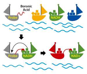
Chang's team was inspired by an existing method of using an enzyme to trigger a reaction between NADH and the probe. Instead of using an enzyme as a catalyst, however, which can limit the technique's application in live cells, the researchers decided to mimic the enzyme's job of 'hooking' NADH and the probe together.
"We added a boronic acid-based function group to the probe, which hooks up with a particular part of the NADH molecule and shortens the distance between them, thereby making the reaction between probe and NADH much easier," explains Chang. "Once the probe and NADH link, the reaction is accelerated between the probe and the compound nicotinamide in the NADH, which then turns on a strong, stable fluorescent signal."
Crucially, NAD+ does not have the same nicotinamide compound, meaning it cannot react with the probe. This allows the researchers to specifically trace NADH, and not NAD+, in living cells.
Once the probe was designed, the team had a second challenge to overcome—it would only work in alkaline environments, so would not function correctly inside living cells, which are pH-neutral. Chang and his team had to modify the boronic acid further so that it would respond to NADH under neutral conditions.
"Our results showed that the turn-on fluorescent probe shows remarkable sensitivity and selectivity to NADH without the need for any additional enzymes," says Chang. This novel method based on imitating enzyme-triggered reactions to design probes could be extended to facilitate the imaging of many other complex biomolecules in the future.
More information: Lu Wang et al. Boronic Acid: A Bio-Inspired Strategy To Increase the Sensitivity and Selectivity of Fluorescent NADH Probe, Journal of the American Chemical Society (2016). DOI: 10.1021/jacs.6b05810
Journal information: Journal of the American Chemical Society


