Microscopic technique to observe antibiotics live in action

A new microscopic technique is enabling scientists to observe the antibiotic daptomycine live in action. This marks an exciting first, because even though doctors have been prescribing this antibiotic for over a decade, its precise mechanisms have remained unclear. Researchers at the University of Amsterdam (UvA), Bonn University and Ruhr University Bochum have now described this mechanism in the forthcoming issue of PNAS.
Antibiotic resistance is a serious threat to public health. Bacteria are constantly adapting to the antibiotics that we use, and finding new antibiotics is vital, emphasises Leendert Hamoen, professor of General Microbiology and one of the current study's leaders. 'Without antibiotics, we may as well give up doing open-heart surgeries and administering cancer treatments, because most people will simply die of infections, just like in the old days.'
Hamoen and his German colleagues deployed cell biology to develop a way to study the activity of new antibiotics up close. Using this technique, they homed in on daptomycine, a so-called 'last-resort antibiotic' that is prescribed in cases where all other antibiotics fail.
First, the scientists tagged the antibiotic so it would light up under a microscope. Comparable florescent labels were then attached to selected functional proteins in bacteria which they though might be inactivated by the antibiotic. Watching through a microscope, the research team were subsequently able to observe in detail just how the antibiotic killed the bacteria.
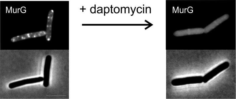
The scientists saw daptomycine clump together at specific regions of the bacterial cell membrane, where they caused a protein needed for cell wall synthesis to disappear from those areas of the membrane. Hamoen explains, 'This means daptomycine drives a vital protein away from the spot where it is active so that the cell wall breaks down and the bacterium dies'. This is the first time that such a mechanism has been observed for antibiotics.
With this new imaging technique, the scientists want to investigate other antibacterial compounds. 'If you want to discover truly effective new antibiotics and chemically enhance them, you've got to know exactly how they work', concludes Hamoen. 'We have now a new tool to study this.'
More information: Anna Müller et al. Daptomycin inhibits cell envelope synthesis by interfering with fluid membrane microdomains, Proceedings of the National Academy of Sciences (2016). DOI: 10.1073/pnas.1611173113
Journal information: Proceedings of the National Academy of Sciences
Provided by University of Amsterdam


