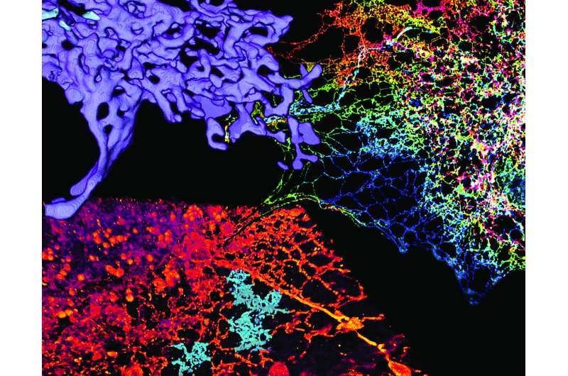
Dense tubular matrices in the peripheral ER. Credit: (c) Science (2016). DOI: 10.1126/science.aaf3928
(Phys.org)—A team of researchers from the U.S. and the U.K. has used high-resolution imaging techniques to get a closer look at the endoplasmic reticulum (ET), a cellular organelle, and in so doing, has found that its structure is not made of tiny sheets of materials, as was thought, but is instead composed of tubule structures. In their paper published in the journal Science, the team describes their research and their theories on why the organelles have such a dynamic structure. Mark Terasaki with the University of Connecticut Health Center offers a Perspective piece providing a short history of such research and outlining the work done by the team in the same journal issue.
Organelles are the structures that reside inside living cells—the ET lies within the cytoplasm of eukaryotic cells and is part of many cellular processes such as protein synthesis, calcium storage, mitochondrial division and lipid synthesis and transfer—because of its tiny size and dynamic nature, it has been difficult to obtain imagery to accurately reveal its structure. In this new effort, the researchers have used a variety of techniques, some cutting edge, to capture the most detailed look at ET to date, thereby overturning some ideas regarding its structure and how it functions.
To capture the images, the team used single-molecule super-resolution techniques along with new ways to illuminate their subject. One, grazing incidence structured illumination microscopy (where light is applied at a perpendicular angle to the target), captured imagery on live cells and provided a major increase in resolution via better lighting and a faster means for capturing images than other techniques, improving resolution in moving objects. Such techniques allowed for capturing sharp images of tubules with diameters as small as 50 nm—each a part of a matrix comprising a network that allows for dynamically responding to the needs of the cell. In the images, the tubules look rather like a large complex of interconnected water pipes.
The researchers suggest the tubule structure allows the ET to adjust very quickly to needs by changing its structure to adapt to cell processes.
More information: J. Nixon-Abell et al. Increased spatiotemporal resolution reveals highly dynamic dense tubular matrices in the peripheral ER, Science (2016). DOI: 10.1126/science.aaf3928
Abstract
The endoplasmic reticulum (ER) is an expansive, membrane-enclosed organelle that plays crucial roles in numerous cellular functions. We used emerging superresolution imaging technologies to clarify the morphology and dynamics of the peripheral ER, which contacts and modulates most other intracellular organelles. Peripheral components of the ER have classically been described as comprising both tubules and flat sheets. We show that this system consists almost exclusively of tubules at varying densities, including structures that we term ER matrices. Conventional optical imaging technologies had led to misidentification of these structures as sheets because of the dense clustering of tubular junctions and a previously uncharacterized rapid form of ER motion. The existence of ER matrices explains previous confounding evidence that had indicated the occurrence of ER "sheet" proliferation after overexpression of tubular junction–forming proteins.
Journal information: Science
© 2016 Phys.org


