Innovative technique to analyse mesoscopic materials directly

Scientists at DESY have developed a method to record the inner structure of individual photonic crystals and similar materials in 3-D. The technique directly reveals the positions of the individual building blocks of a crystal, without the need for assumptions, models or averaging. Photonic crystals have a wide range of applications in information technology, chemistry and physics. The team around main author Ivan Vartaniants from DESY reports their work in the scientific journal Physical Review Letters.
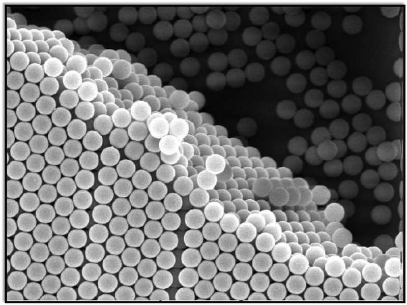
Photonic crystals are composed of particles in the size range of roughly 200 to 400 nanometres. Correspondingly, their inner structure is in the range of the wavelength of visible light. This "submicron" structure enables various effects to manipulate optical photons – hence the name. An example for a natural photonic crystal is the gem opal, consisting of small glass beads of silica gel, measuring 150 to 400 nanometres each. "Also, the colours of butterfly wings are often produced by similar photonic structures," explains Vartaniants.
More generally, the periodic arrangements of small particles are called colloidal crystals. The characteristics of colloidal crystals strongly depend on their exact inner structure. "For many applications it is vital to understand the real structure of colloidal crystals in every detail," says co-author Andrei Petukhov from Utrecht University in the Netherlands. For instance, to use them as waveguides in optical information networks it is often crucial to introduce well-defined defects in the crystal structure.

"However, analysing these structures proved to be complicated," says Vartaniants. "Optical microscopy often does not deliver the necessary spatial resolution. Electron microscopy can see much more details but, since electrons do not penetrate far below the surface, the bulk of the crystal remains hidden. X-rays easily traverse the whole crystal and can reveal fine details, but they do not image the crystal, its structure has to be calculated from the way the X-rays are scattered by the sample, usually needing assumptions or models for the structure under investigation."
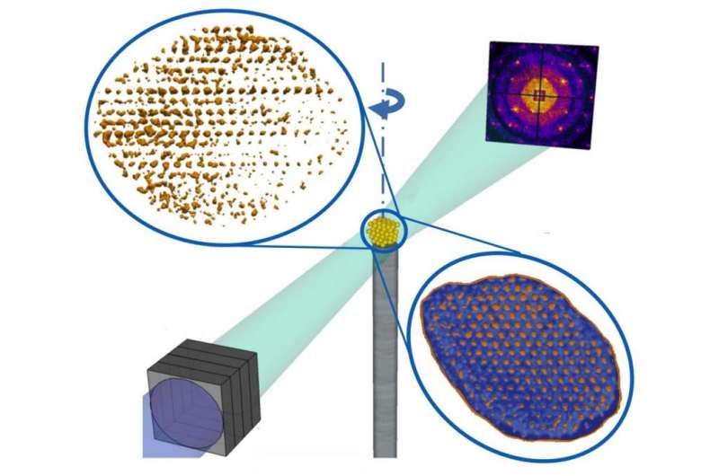
The team used the brilliant, coherent X-rays from DESY's research light source PETRA III to investigate the inner structure of an artificially produced colloidal crystal from Petukhov's lab in Utrecht, grown from silica spheres with a diameter of 230 nanometres each. "The small investigated crystal measured two by three by four micrometres," says co-author Michael Sprung, head of the DESY beamline P10 where the investigation was performed. A micrometre is 1000 nanometres. "The whole crystal was illuminated by the coherent X-rays from PETRA III and rotated to record the structure from every side."
Like in conventional crystallography, the X-rays are diffracted by the crystal lattice, producing a characteristic diffraction pattern on the detector from which the inner structure can be calculated. "But while conventional crystallography concentrates on the positions of bright spots in the diffraction pattern, known as Bragg peaks, the coherent, laser-like illumination of the sample also produces interference patterns in between the Bragg peaks," explains first author Anatoly Shabalin from DESY. Using this information, the scientists could determine not just the general inner structure of the crystal, but the positions of the individual silica spheres throughout their sample. This analysis directly revealed the individual, three-dimensional inner structure of the crystal, including defects of different types.
"Our method opens up new ways to visualise the inner three-dimensional structure of mesoscopic materials like photonic crystals with coherent X-rays," says Vartaniants. "Especially with the next generation of X-ray light sources, this technique can pave the way to analyse individual nanocrystals with atomic resolution."
More information: A. G. Shabalin et al. Revealing Three-Dimensional Structure of an Individual Colloidal Crystal Grain by Coherent X-Ray Diffractive Imaging, Physical Review Letters (2016). DOI: 10.1103/PhysRevLett.117.138002
Journal information: Physical Review Letters
Provided by Helmholtz Association of German Research Centres


