Combined approach used to investigate photophysics of green fluorescent protein

A substance squeezed from a jellyfish found off the western coast of North America has transformed modern cellular biology. The substance is green fluorescent protein (GFP), which, when added to viruses and proteins, brings their life and function into view. Now, by combining experiment, computation, and theory, scientists at Pacific Northwest National Laboratory and colleagues at Louisiana State University have discovered new insights about GFP's behavior. This in turn can lead to even more ways to exploit this valuable monitor of biological processes.
By adding a few water molecules to the GFP chromophore—the part of the molecule responsible for its color—the scientists simulated crystallographic water molecules in GFPs. When water is added, the excited state that generates fluorescence is more stable. Water apparently shuts the channel to electron emissions, effectively shutting off electron autodetachment competition and allowing fluorescence.
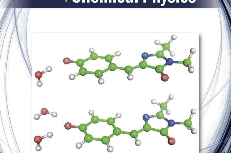
The scientists' findings provide a more accurate understanding of this extremely useful protein. The two water molecules shown on the December 14 cover of The Journal of Chemical Physics make a huge difference in the behavior of the GFP. In turn, this can lead to eventual control and manipulation of processes where GFP is used.
GFPs (see sidebar) are invaluable as markers for monitoring processes in living cells, such as in cancer research, where GFP-labeled cells model how cancer spreads to organs. Their versatility and value has led to the synthesis of new types of GFPs with different colors and new classes of photoactivatable fluorescent proteins for use in ultra-resolution imaging and optical data storage of raw data images from living cells and tissues.
The photophysics of GFP and its chromophore depends on its local structure and environment. Despite extensive experimental and computational studies, many open questions remain about the key fundamental variables governing this process.
One question is what controls the efficiency of light emission. When GFP absorbs light, only some of the energy is converted into a fluorescent signal, with the rest being lost by a process known as relaxation or deactivation. The fraction of energy emitted as fluorescence versus that lost by relaxation dictates GFP's efficiency. What the PNNL and LSU scientists wanted to know was how the protein's internal environment—in this case, water—affects specific types of relaxation.
"We know the relaxation that competes against the fluorescence is critically dependent on the GFP chromophore's local environments, but we don't fully understand the details of why it happens," said PNNL chemical physicist Dr. Xue-Bin Wang, senior author of the article. "If we get rid of surrounding molecules by using the gas phase, the fluorescence goes away. And in solution, no fluorescence occurs at room temperature. But fluorescence returns at low temperatures. Is it caused by the intrinsic electronic structure properties of the chromophore in the protein? Or is it caused by other molecules in its environment? Our results showed it was the latter."
GFP is centrally important to modern cell biology. Its discovery and development garnered the 2008 Nobel Prize in Chemistry for scientists Osamu Shimomura, Martin Chalfie, and Roger Tsien. Gaining a fundamental molecular understanding of how GFP works can lead to the ability to control, engineer, or manipulate systems for new applications, such as biosensors, or for advanced imaging.

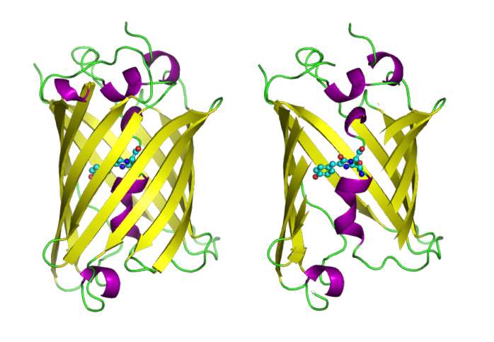
In the cartoons shown here, the GFP molecules are shown in full, and with the side of the surrounding barrel cut away (right) to reveal the chromophore, which is highlighted as a ball-and-stick model. Fluorescence arises when a molecule absorbs certain colors of visible light and then reemits a different, lower energy color.
The interactions of the chromophore with its surroundings will dramatically affect the fluorescent properties of GFP, such as the color or intensity of the fluorescence. Researchers are currently trying to understand the underlying physics of these processes.
By adding water and comparing the computation with experimental results, the scientists could determine that their theory of the molecules' structure was accurate.
The team combined negative ion photoelectron spectroscopy (NIPES) with theoretical calculations to create a probe to identify the exact conformers of clusters of the p-hydroxybenzylidene-2,3-dimethylimidazolinone anion (HBDI-), a model of the GFP chromophore. They used NWChem, an open-source computational chemistry software package supported by the Department of Energy (DOE) Office of Biological and Environmental Research (BER) and developed at PNNL with unique capabilities regarding excited states and structure characterization, and the supercomputer Cascade, at EMSL.
In a previous study by Wang and collaborators at the Chinese Academy of Sciences, the GFP chromophore itself was studied in the gas phase. In the current study, the scientists took another step farther by adding the water molecules. The notable addition was the use of advanced computer simulation techniques developed in collaboration with LSU.
"In the current paper, the important component was coming up with an accurate theory. In an experiment, when we obtain a signal, we don't 'see' what is happening. Computer simulations using NWChem and EMSL supercomputing resources, give us the necessary details," said co-author Dr. Karol Kowalski.
"It's a puzzle," said computational scientist Dr. Marat Valiev, one of the co-authors. "You can't interrogate the protein system as a whole to obtain key molecular-level parameters governing photoresponses of GFP. We have to disassemble it piece by piece, examine each piece, and then put it back together, which is best approached through combining experiment and simulation."
The scientists showed that the first few water molecules progressively stabilize the excited state of the chromophore. "This could be an important role of water molecules in GFPs that has not yet been fully explored," said Wang.
More information: S. H. M. Deng et al. Vibrationally Resolved Photoelectron Spectroscopy of the Model GFP Chromophore Anion Revealing the Photoexcited S State Being Both Vertically and Adiabatically Bound against the Photodetached D Continuum , The Journal of Physical Chemistry Letters (2014). DOI: 10.1021/jz500869b
Kiran Bhaskaran-Nair et al. Probing microhydration effect on the electronic structure of the GFP chromophore anion: Photoelectron spectroscopy and theoretical investigations, The Journal of Chemical Physics (2015). DOI: 10.1063/1.4936252
Journal information: Journal of Chemical Physics , Journal of Physical Chemistry Letters
Provided by Pacific Northwest National Laboratory


