Green tea's popularity has grown quickly in recent years. Its fans can drink it, enjoy its flavor in their ice cream and slather it on their skin with lotions infused with it. Now, the tea could have a new, unexpected role—to improve the image quality of MRIs. Scientists report in the journal ACS Applied Materials & Interfaces that they successfully used compounds from green tea to help image cancer tumors in mice.
Sanjay Mathur and colleagues note that recent research has revealed the potential usefulness of nanoparticles—iron oxide in particular—to make biomedical imaging better. But the nanoparticles have their disadvantages. They tend to cluster together easily and need help getting to their destinations in the body. To address these issues, researchers have recently tried attaching natural nutrients to the nanoparticles. Mathur's team wanted to see if compounds from green tea, which research suggests has anticancer and anti-inflammatory properties, could play this role.
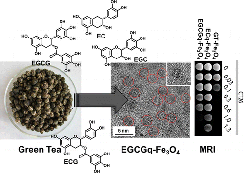
Using a simple, one-step process, the researchers coated iron-oxide nanoparticles with green-tea compounds called catechins and administered them to mice with cancer. MRIs demonstrated that the novel imaging agents gathered in tumor cells and showed a strong contrast from surrounding non-tumor cells. The researchers conclude that the catechin-coated nanoparticles are promising candidates for use in MRIs and related applications.
More information: Enhanced In Vitro and In Vivo Cellular Imaging with Green Tea Coated Water-Soluble Iron Oxide Nanocrystals, ACS Appl. Mater. Interfaces, Article ASAP. DOI: 10.1021/am508404t
Abstract
Fully green and facile redox chemistry involving reduction of colloidal iron hydroxide (Fe(OH)3) through green tea (GT) polyphenols produced water-soluble Fe3O4 nanocrystals coated with GT extracts namely epigallocatechin gallate (EGCG) and epicatechin (EC). Electron donating polyphenols stoichiometrically reduced Fe3+ ions into Fe2+ ions resulting in the formation of magnetite (Fe3O4) nanoparticles and corresponding oxidized products (semiquinones and quinones) that simultaneously served as efficient surface chelators for the Fe3O4 nanoparticles making them dispersible and stable in water, PBS, and cell culture medium for extended time periods. As-formed iron oxide nanoparticles (2.5–6 nm) displayed high crystallinity and saturation magnetization as well as high relaxivity ratios manifested in strong contrast enhancement observed in T2-weighted images. Potential of green tea-coated superparamagnetic iron oxide nanocrystals (SPIONs) as superior negative contrast agents was confirmed by in vitro and in vivo experiments. Primary human macrophages (J774A.1) and colon cancer cells (CT26) were chosen to assess cytotoxicity and cellular uptake of GT-, EGCGq-, and ECq-coated Fe3O4 nanoparticles, which showed high uptake efficiencies by J774A.1 and CT26 cells without any additional transfection agent. Furthermore, the in vivo accumulation characteristics of GT-coated Fe3O4 nanoparticles were similar to those observed in clinical studies of SPIONs with comparable accumulation in epidermoid cancer-xenograft bearing mice. Given their promising transport and uptake characteristics and new surface chemistry, GT-SPIONs conjugates can be applied for multimodal imaging and therapeutic applications by anchoring further functionalities.
Journal information: ACS Applied Materials and Interfaces
Provided by American Chemical Society


