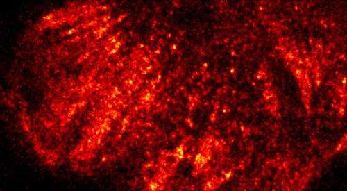
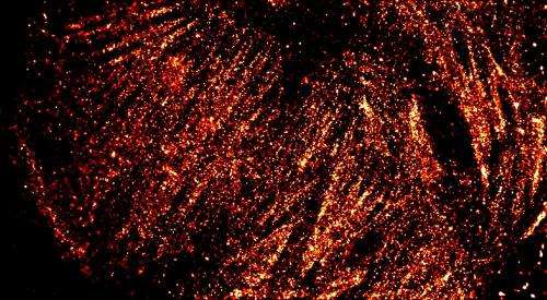
Standard ensemble of the actin cytoskeleton, structures in the cell thought to be important to the protein-protein interaction implicated in many cancers. Credit: STFC
A revolutionary new £1M laser instrument in the UK is set to illuminate the innermost workings of DNA, protein, enzyme and other molecules that play a critical role in key natural processes in humans, animals and plants. Using synchronised ultrafast infrared lasers to explore how biomolecules react and interact when struck by light, LIFEtime will provide the basis for developing better medicines, innovative cell-imaging techniques and tiny biomolecular 'probes' that can be inserted into cells to gather information.
The instrument is due to be fully operational shortly and is based at the Science and Technology Facilities Council's (STFC) Central Laser Facility (CLF). Developed with Biotechnology and Biological Sciences Research Council (BBSRC) support, LIFEtime's uniqueness lies in its ability to measure how a biomolecule responds to a laser flash within the first trillionth of a second and to assess follow-on reactions occurring on timescales of a thousandth of a second or more. This remarkable 'two in one' capability saves time and money, and minimises potential damage to the samples being examined.
Key to establishing the technology as a useable tool is the development and application of computational capabilities that can analyse and interpret the high volume, highly complex datasets generated by LIFEtime.
Professor Tony Parker of the CLF explains: "We're combining complementary data obtained from crystal samples by the Diamond Light Source, based next door, and using this to develop computer code that provides a structural identification tool for solution studies - as real biology works in our cells in our lifetime."
Professor Steve Meech of the University of East Anglia will use LIFEtime to study the dynamics of photosensitive proteins. He says: "We need extremely sensitive measurements as we're looking for small changes and want to track responses from the laser's first interaction with the protein right through to milliseconds afterwards. The LIFEtime apparatus with its high stability and high repetition rate will allow us to record these changes and help us take a significant step forward in understanding protein function."
-
Super-resolution image of the actin cytoskeleton, structures in the cell thought to be important to the protein-protein interaction implicated in many cancers. This is from STORM(STochastic Optical Reconstruction Microscopy)another super-resolution method used in the Central Laser Facility, showing the difference high resolution imaging can make to the level of detail. Credit: STFC
-
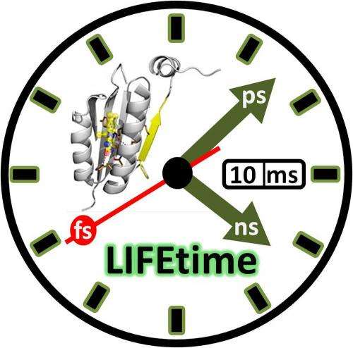
A representation of the times scales over which LIFEtime can probe using clock hands representing femtoseconds, picoseconds and nanoseconds and calendar representing the “slow millisecond” timescale. Credit: Paul Donaldson
Provided by Science and Technology Facilities Council


