Artificial intelligence annotates medical images

Biomedical research students at the University of Sydney have joined international efforts to improve the automatic interpretation of 3D anatomical images used by health practitioners.
The team has received international recognition for their unique algorithm that automatically analyses three-dimensional computed tomographic (3D CT) liver images. The team's work was awarded first place in the annual Cross Language Evaluation Forum image challenge. The detailed results will be presented at the Cross Language Evaluation Forum (CLEF) to be held in Sheffield in the United Kingdom.
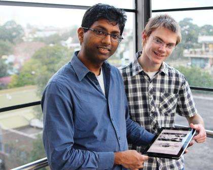
ImageCLEF hosts a variety of challenges. This year the aim of their medical imaging challenge was to automatically annotate 3D CT images of the liver based on an analysis of the images says team member Dr Ashnil Kumar, a post-doctoral fellow in the University's School of Information Technologies.
"Medical imaging is now a fundamental aspect of healthcare delivery but the challenge facing clinicians is how best to extract or identify relevant information from these massive data sets," states Dr Kumar.
"Subtle differences in medical images are often critical in determining patient outcomes. Our work is part of a long-term worldwide goal to develop better clinical support technologies.
"Advances in imaging technology means that we now have bigger, better images, in 3D for example that can be used to detect these differences. The downside is the increased time and effort needed by expert radiologists to analyse them."
A major challenge is developing technologies to better support a radiologist's workflow. The ability to automatically analyse and annotate images, and then generate a structured report from the analysis and annotations has the potential to improve the efficiency of the radiologist's practice.
Ideally, this would have flow on effects in terms of patient throughput and faster communication between radiologists, oncologists, and patients.
Our aim for the future is to build smarter, more accurate systems that enable clinical staff to work more efficiently. This project is the first step in this in goal.
Electrical engineering student, Shane Dyer who was central to the team's efforts says:
"We know that analysis of 3D liver scans is a time consuming task for physicians, but our team knows we can create technology to overcome.
"We created a system that uses artificial intelligence (AI) techniques in order to automatically determine various properties of a liver for example number of lesions, size of the liver.
"Future work could include detecting when a property is abnormal, and the implementation of such a system could have the flow-on effect of reduced patient waiting times.
Essentially, our challenge was to create AI that could fill in a form similar to a radiologist's report. Examples of the annotation included liver and tumour properties such as shape, location, calcification, interaction with nearby vessels," says Shane.
Provided by University of Sydney


