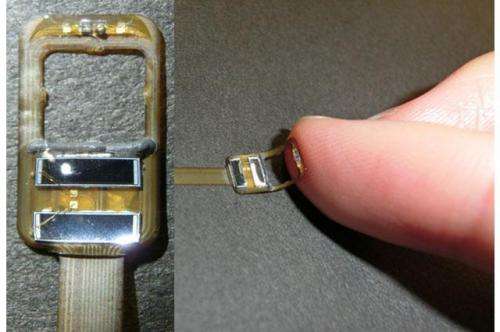
The miniaturised optical probe used in the work; made using flexible printed circuit technology.
Research from Japan has produced a NIRS probe small enough for implanted use in brain functional imaging of humans. Based on flexible printed circuit technology, the probe is also cheaper and simpler to produce than conventional micro fiber optics devices.
Imaging activity
Near infrared spectroscopy (NIRS) is a spectroscopic method for measuring blood volume and oxygen saturation in both brain and muscle tissue by the light absorption of oxyhaemoglobin (O2Hb) and deoxyhaemoglobin (HHb). Haemoglobin concentration and oxygenation are useful as indicators of activity within tissue and its metabolism; this is useful in a wide range of medical applications including the examination of muscles in sports medicine and examining brain function by using NIRS to detect and map active areas of the brain.
There are several types of NIRS systems currently in use. Single-point systems can measure changes in concentration of O2Hb and HHb. Spatially-resolved NIRS, time-resolved NIRS and phase-modulated NIRS can measure absolute values of concentration of haemoglobin, oxygen saturation and blood volume. Brian functional imaging by NIRS is made possible by multichannel NIRS systems.
NIRS probes need to incorporate both a light source and sensors to read the way the light interacts with the tissue. The optical probes of existing NIRS systems are designed for non-invasive use, attaching to the outside of the body. Current commercially available probes are typically around 10 x 30 mm in area and 5 mm thick. With probe volumes in the region of 1500 mm3, they are not suited to implanted use or for measuring activity in the brains of small animals or use in evaluating the epileptogenic focus in the cerebral cortex.
Small volume, high volume
The probe design used in the work from Shizuoka University is 100 times smaller than these conventional NIRS probes with a volume of only 15 mm3. The new design is also easier to manufacture than alternatives as it is fabricated using flexible printed circuit (FPC) technology rather than using micro fibre optics, as author Dr Masatsugu Niwayama explained: "When using micro fibre optics to manufacture implantation probes, expert techniques are required to connect two fibre cores. Fragility of the fibre also increases the difficulty of probe assembly. Recently, FPC substrate with wire bonding can be ordered from many printed circuit board companies without much difficulty. The easy manufacturing would enable us to mass produce the probes and even make them disposable items. Disposable probes will be effective to help prevent infection in hospitals."
The team have previously developed a similar size of probe that was only able to measure changes in haemoglobin concentration and had the problem that the measurement sensitivity of each tissue type was unclear quantitatively. In the work presented in their Letter they have addressed both of these issues, presenting absolute value measurements of haemoglobin concentration based on theoretical sensitivity analysis and a spatially resolved method. They also present analysis of the difference in measurement sensitivities for the brain and scalp between implanted sensors and body surface sensors.
Non-invasive future
Since completing the reported work the Shizuoka team have already conducted some in vivo tests with animals and are confident that use in humans should follow within a year for acute (short term) NIRS-ECoG (electrocorticography) simultaneous measurement in patients during neurosurgery. The key hurdle remaining to clear the probe for chronic (long term) implantation in human subjects is completion of toxicity/biocompatibility tests under chronic implantation in large animals to prove the safety and reliability of the device for extended use.
Meanwhile, the team are working on increasing the NIRS recording channels to observe the localised/distributed hemodynamic activity related to brain diseases. "Our interests are now moving into revealing the connectivity between neuronal electrophysiological activity and the focal hemodynamics, and its difference between the normal and abnormal (diseased) brain. We also hope that our method will solve other remaining problems of clinical NIRS application, such as artefacts due to scalp blood flow," said Niwayama.
"Recently a small number of researchers have been trying to observe the cortical hemodynamics directly from the brain surface for high accuracy recording, and our device can facilitate the spread of this experimental method since the measurement system can be manufactured less expensively than optical-fibre-based products." The team's hope is that in the next ten years work like theirs will enable new findings in cortical hemodynamics that will then see the use of non-invasive NIRS equipment become widespread in the diagnosis of brain functional diseases.
More information: "Implantable thin NIRS probe design and sensitivity distribution analysis." M. Niwayama, T. Yamakawa. Electronics Letters, Volume 50, Issue 5, 27 February 2014, p. 346 – 348. DOI: 10.1049/el.2013.3921 , Print ISSN 0013-5194, Online ISSN 1350-911X
Journal information: Electronics Letters
Provided by Institution of Engineering and Technology


