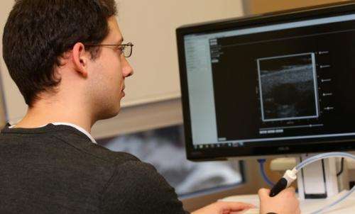
Ultra-small ultrasounds

Andre Bezanson is still working on his PhD, but he's already making waves in the field of ultrasound imaging.
Bezanson, a biomedical engineering student, is conducting research that allows scientists and clinical researchers to image smaller objects with a higher resolution using ultrasound technology—and at a lower cost than with traditional high frequency ultrasound devices.
His work recently earned him the Mitacs Award for Outstanding Innovation, an award given annually to five Mitacs-sponsored innovators chosen out of thousands of nominees from across Canada. Bezanson was also a recipient of Dalhousie's Marble Prize and an attendee at the 2013 IEEE Ultrasonics, Ferroelectrics, and Frequency Control Society conference in Prague, where he was honored with the Student Paper Competition Award.
Mitacs is a Canadian not-for-profit organization that offers funding for internships and fellowships at Canadian universities for international undergraduate and graduate students.
"I did a work term where I had additional funding through Mitacs," Bezanson says. "I partnered up with Daxsonics, which is a startup ultrasound company here in Halifax. During the work term we focused on producing a high resolution ultrasound system primarily focused on scanning small animals."
A practical solution
The device that Bezanson and Daxsonics came up with is different than other high resolution ultrasound systems because it replaces expensive and bulky electromagnetic motors with a piezoelectric bimorph—a simple, credit card-shaped object that bends under an electrical field and costs only about $20.
He says the primary application for the system is preclinical studies with small animals.
"There are no low cost systems out there with a price-point that makes the unit accessible to a standard academic researcher, so we were looking to bring the cost down to the point where academic researchers can afford the systems on a typical grant," he says. "We were able to pull that off during my work term, which was a massive breakthrough."
Many labs that are unable to afford an imaging device for small animals must breed large sample populations and employ dissection as a primary imaging technique. With an affordable ultrasound device, labs can dramatically cut down on the sample size needed and non-invasively track physiological changes, such as tumor growth, directly. This saves researchers time and money, and saves the lives of many lab animals as well.
Though it's currently being viewed as a pre-clinical research tool, the device may be used in other research labs such as biology or neuroscience. "The more people who get to use it, the more people coming out with interesting, relevant data," Bezanson enthuses.
Provided by Dalhousie University


