New modeling tool developed for imaging atomic displacement in crystals

(Phys.org) —You use crystals everyday: sugar in your coffee, the active ingredient in hand warmers, maybe a diamond stud in your ear.
A crystal is built of atoms arranged in a repeat pattern in all three dimensions. X-rays are good at detecting this pattern because x-rays can only diffract into specific directions that depend on the symmetry and repeat distances in the pattern. If the atoms are displaced because of strain, their pattern of displacement can be calculated from the directional change of the diffracted x-ray beam. That is the simple theory.
Now think of an ocean wave hitting the piles of a jetty. If the wave hits once and leaves the piles, that's an example of kinematical diffraction. Next picture the wave staying with the piles and hitting the piles multiple times, which causes a complex pattern of ripples. That's dynamical diffraction. It usually occurs in single crystals over a micron in size and results in phenomena that can't be explained by the simpler kinematical diffraction theory.
The dynamical diffraction with "multiple hits" is obviously much harder to model in diffraction theory. For this reason, all existing dynamical approaches are usually applicable only to simple crystal geometries, for example, by assuming an infinitely large crystal block.
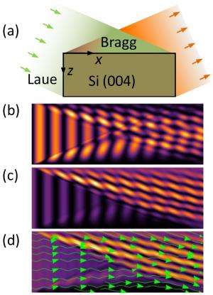
At the National Synchrotron Light Source II (NSLS-II), now under construction at Brookhaven National Laboratory, physicists Hanfei Yan and Li Li have developed an ingenious method to model x-ray dynamical diffraction for any crystal size and geometry. Their theoretical model opens up opportunities to explore new ways of imaging strain fields – a quantitative measure of the relative atomic displacement – in crystals, particularly in microcrystals, which are important to nanotechnology applications. It also sheds light on many fundamental diffraction problems that have not been fully understood before.
Yan and Li are part of the team building the Hard X-ray Nanoprobe (HXN) beamline, one of an initial suite of beamlines that will be ready for early science commissioning experiments in the fall of 2014 at NSLS-II.
According to Yan, x-ray dynamical diffraction is poised to become a very important modeling tool for HXN because strain can affect optical, mechanical and electrical properties of crystalline materials. Knowing how the strain distributes is vital to understanding the underlying physics. A nanobeam will enable the strain imaging at the nanoscale, which is from 1 to 100 nanometers (a strand of human DNA is from 1.8 to 2.3 nanometers in diameter).
"Dynamical diffraction theory is fundamental in x-ray scattering science," said Yan. "Our theoretical development will enable scientists to correctly image how the atomic displacement changes in micron-size single crystals at spatial resolutions ranging from 10 to 30 nanometers, with the long-range goal of one nanometer at HXN."
In the past, various nanodiffraction measurements have been made to probe strain at the nanoscale. "Without a rigorous data analysis approach, dynamical artifacts can arise and lead to erroneous results," Li added. "Our method provides a solution to such problems."
In further development of their theory, the scientists will incorporate a nanobeam into the model to develop the data-analysis method for quantitative strain imaging at a high spatial resolution. They also plan to extend a newly emerged technique known as Bragg coherent diffractive imaging into the dynamical diffraction regime.
Li is also developing PyLight, a data analysis framework for NSLS-II, in collaboration with Dantong Yu in Brookhaven's Computational Science Center (CSC) and other Brookhaven scientists. "Our new modeling approach will be incorporated into this software framework to enable more scientists to share the strain-analysis software and handle much larger datasets with CSC's high-performance computing clusters," Li said.
Yan and Li published their theoretical method in Physical Review B, January 2014. Their work was supported by the U.S. Department of Energy.
More information: Science Paper: X-ray dynamical diffraction from single crystals with arbitrary shape and strain field: A universal approach to modeling: prb.aps.org/abstract/PRB/v89/i1/e014104
Journal information: Physical Review B
Provided by Brookhaven National Laboratory


