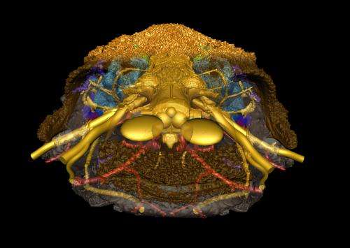
This image shows a reconstruction in three dimensions of the skull of the small fossil fish Romundina (415 million years old) that was scanned at the ESRF. The internal structures of the face reveal the internal anatomy show a mixture of structures of jawless and jawed vertebrates (in anterior view). External bones of two different kinds in orange and pink grey, nerves and cranial cavity in yellow, arteries in red, veins in dark blue and inner ears in light blue; anterior part of the bone rendered semitransparent. Credit: Vincent Dupret, Uppsala University
A team of French and Swedish researchers have presented new fossil evidence for the origin of one of the most important and emotionally significant parts of our anatomy: the face. Using micron resolution X-ray imaging, they show how a series of fossils, with a 410 million year old armoured fish called Romundina at its centre, documents the step-by-step assembly of the face during the evolutionary transition from jawless to jawed vertebrates. The research is published in Nature on February 12, 2014.
Vertebrates, or backboned animals, come in two basic models: jawless and jawed. Today, the only jawless vertebrates are lampreys and hagfishes, whereas jawed vertebrates number more than fifty thousand species, including ourselves. It is known that jawed vertebrates evolved from jawless ones, a dramatic anatomical transformation that effectively turned the face inside out.
In embryos of jawless vertebrates, blocks of tissue grow forward on either side of the brain, meeting in the midline at the front to create a big upper lip surrounding a single midline "nostril" that lies just in front of the eyes. In jawed vertebrates, this same tissue grows forward in the midline under the brain, pushing between the left and right nasal sacs which open separately to the outside. This is why our face has two nostrils rather than a single big hole in the middle. The front part of the brain is also much longer in jawed vertebrates, with the result that our nose is positioned at the front of the face rather than far back between our eyes.
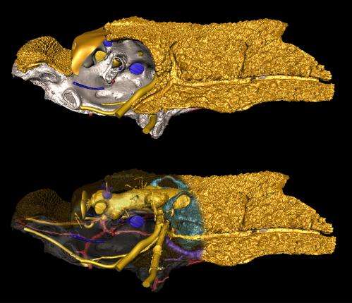
These two images show the skull of the small fossil fish Romundina (415 million years old) scanned at the ESRF and digitally reconstructed in three dimensions. The internal structures of the face reveal the internal anatomy show a mixture of structures of jawless and jawed vertebrates (here in left lateral view, top). External bones of two different kinds in orange and pink grey, nerves and cranial cavity in yellow, arteries in red, veins in dark blue and inner ears in light blue; anterior part of the bone rendered semitransparent in bottom image (bottom). Credit: Vincent Dupret, Uppsala University
Until now, very little has been known about the intermediate steps of this strange transformation. The scientists studied the skull of Romundina, an early armoured fish with jaws, or placoderm, from arctic Canada. The skull is part of a collection of the French National Natural History Museum in Paris.
Romundina has separate left and right nostrils, but they sit far back, behind an upper lip like that of a jawless vertebrate. "This skull is a mix of primitive and modern features, making it an invaluable intermediate fossil between jawless and jawed vertebrates", says Vincent Dupret of Uppsala University, one of two lead authors of the study.
In this video sequence, different transparencies provide an ensemble view of the blood vessel network (red: arteries; blue: veins), nerves and endocranial cavity (yellow), shape of telencephalon, nasal sacs and rostronasal capsule, notochord (maroon), and inner ears (light blue) of the fish fossil Romundina. Credit: Vincent Dupret, Uppsala University and Philippe Loubry, CNRS-MNHN,
By imaging the internal structure of the skull using high-energy X-rays at the European Synchrotron (ESRF) in Grenoble, France, the authors show that the skull housed a brain with a short front end, very similar to that of a jawless vertebrate.
"In effect, Romundina has the construction of a jawed vertebrate but the proportions of a jawless one", says Per Ahlberg, of Uppsala University and the other lead author of the study. "This shows us that the organization of the major tissue blocks was the first thing to change, and that the shape of the head caught up afterwards", he adds.
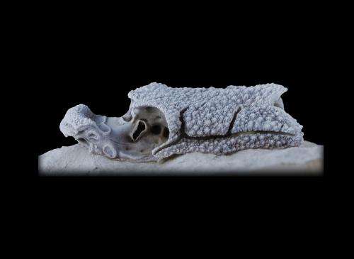
The skull of the small fossil fish Romundina (415 million years old). photo of specimen in lateral view. Credit: Philippe Loubry, CNRS-MNHN
By placing Romundina in a sequence of other fossil fishes, some more primitive and some more advanced, the authors were able to map out all the main steps of the transition.
"Without the intense X-rays produced at the ESRF, we would not have been able to create a virtual representation of the internal structures of the skull" said Sophie Sanchez from The European Synchrotron (ESRF) in Grenoble.
Journal information: Nature
Provided by European Synchrotron Radiation Facility


.jpg)


