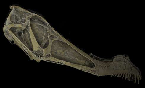
Museum CT scan of a 100-million-year-old pterosaur, species Anhanguera.
(Phys.org) —The inside of the skull of a 100-million-year-old pterosaur has been seen by Natural History Museum fossil experts for the first time. Computed tomography (CT) scans revealed details of the ancient flying reptile's braincase that will help scientists discover more about its behaviour.
The skull belongs to the extinct species Anhanguera - an Early Cretaceous fish-eating pterodactyloid with a long snout and a wingspan of 4-5 metres. The fossil skull, uncovered in Brazil, is half a metre long and is displayed in the Museum - the only Anhanguera fossil on public display in the UK.
Preparing fossils for study and display
To prepare the Anhanguera specimen for study, it went through a 2-year acid preparation process of being immersed in dilute acetic acid to dissolve away the limestone rock surrounding the skull. This was done before it was scanned, in fact before the CT-scan technology was even available to the Museum scientists.
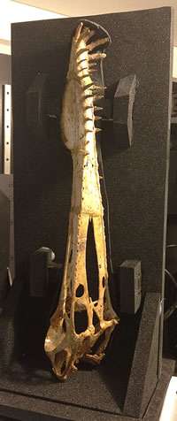
The half a metre long Anhanguera fossil on its special mount to be CT scanned. It had to be scanned upright and rotated 360 degrees.
Museum pterosaur and crocodile curator Lorna Steel explains, 'This takes a long time and the specimen has to be lifted out of the acid periodically, and washed in running water, to wash away all of the acid, and then dried.'
'A protective coating is then applied to the exposed bones, to protect them from the next immersion in acid.'
Scanning the fossil
Scanning the fossil skull was a delicate process. Because it was so long, it could not be placed into the CT scan flat and it needed to stand upright, supported by a special mount made of materials that allowed the scan rays to pass through it. The fossil needed to be able to rotate 360 degrees.
The Museum's conservation team are used to these tricky manoeuvres as they deal with a huge variety of specimens, including some of the around 1000 pterosaur fossils looked after at the Museum.
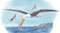
Illustration of the ancient flying reptile, Anhanguera. It had a 4-5m wingspan. Credit: Anness Publishing Ltd / Natural History Museum, London
'The Anhanguera is the largest dinosaur fossil I have scanned,' says Dr Farah Ahmed, Museum CT Facility Manager and Specialist. 'It had to be scanned in two halves then stitched together using computer software which was quite tricky'. It took a couple of days to produce the final images'.
Braincase details revealed
The images produced from the scans reveal fine details of the inside of the braincase - something that otherwise cannot be seen and that will provide valuable information for scientists studying these ancient creatures.
'The scan results can tell us about the shape of the brain,' says Steel. 'This in turn shows which parts of the brain were well-developed or not, and so we can tell which senses eg vision, smell, hearing, balance, were important in the animal.' For example, scientists used CT scans to discover that the earliest known bird, Archaeopteryx, had hearing similar to an emu.
'Researchers could also model the mechanical properties of the skull,' adds Steel. Other CT scan studies have allowed scientists to discover how Diplodocus combed and raked leaves from branches, and that some ancient crocodiles ate like today's killer whales.
Another important use of the scanned digital specimen is that it can be made available to researchers worldwide more easily without the need to transport or handle the precious object.
Other scans
Other recent scans the Museum team have carried out for researchers include the snout of another pterosaur, Istiodactylus, from the Isle of Wight that is about 115-120 million years old to study snout structure. And a Jurassic fossil crocodile called Pelagosaurus, from France, for a study into the evolutionary history of crocodiles.
The Anhanguera skull CT scan data is being studied by USA researchers and they hope to present their results in May. So while we wait for the outcome, its fossil is back on display in the From the Beginning gallery to be enjoyed by the millions of visitors to the Museum who pass through each year.
Provided by Natural History Museum


