Biodiversity exploration in the 3-D era

Taxonomy – the discipline that defines and names groups of organisms – is a field of science that still employs many of the methods used during the beginnings of the discipline in the 18th century. Despite the increasing use of molecular information to delineate new species, the study of the morphology of specimens remains one of the major tasks of taxonomists. These studies often require first-hand examination of the reference specimens (so-called type material) deposited at museum collections around the globe - a time-consuming and laborious task.
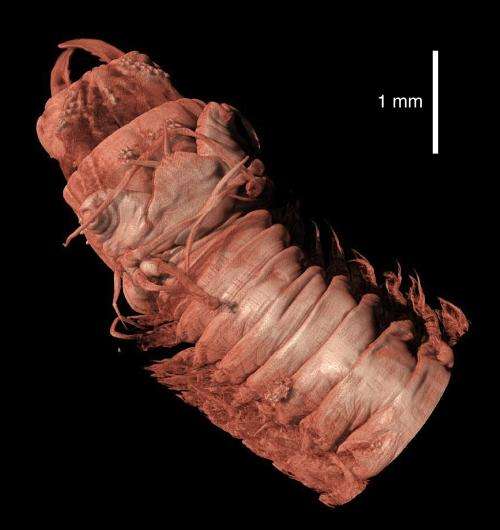
To facilitate this procedure, a group of researchers from the Hellenic Centre for Marine Research (HCMR) are exploring the possibilities offered by 3D digital imaging. In a recent article published in the open-access journal ZooKeys, the researchers use X-ray computed tomography to create digital, three-dimensional representations of tiny animals, displaying both internal and external characteristics of the specimens at a detail level similar to that of the microscope.
To demonstrate their method, the researchers imaged a number of polychaete species (marine bristle-worms)—the choice of this group being obvious to Sarah Faulwetter, the leading author, because "despite being ecologically very important, these animals exhibit a fascinating diversity of forms and tissue types, allowing to test the methodology across a range of samples with different characteristics".
The resulting interactive 3D models allow any researcher to virtually rotate, magnify or even dissect the specimen and thus extracting new scientific information, whereas the structure and genetic material of the analysed specimen are kept intact for future studies.

The team stress the importance of 3D imaging methods for taxonomy on its way into the twenty-first century: "Our vision for the future is to provide a digital representation of each museum specimen, simultaneously accessible via the internet by researchers and nature enthusiasts worldwide," says the team leader, Dr Christos Arvanitidis from HCMR.
The instant accessibility of specimens will speed up the creation and dissemination of knowledge. As the authors point out, "human efforts, combined with novel technologies, will help taxonomy to turn into a cyberscience whose discoveries might rival those made during the great naturalist era of the nineteenth century."
More information: Faulwetter S, Vasileiadou A, Kouratoras M, Dailianis T, Arvanitidis C (2013) Micro-computed tomography: Introducing new dimensions to taxonomy. ZooKeys 263: 1-45. doi: 10.3897/zookeys.263.4261
Journal information: ZooKeys
Provided by Pensoft Publishers


.jpg)


