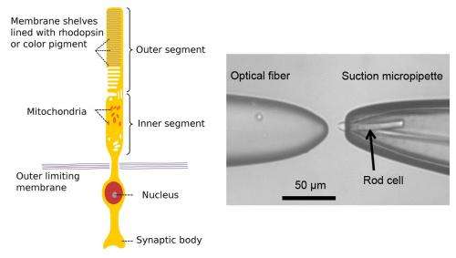
(Left) Structure of a rod cell in the retina. Photons absorbed in the outer segment cause a rearrangement of rhodopsin molecules, which initiates a sequence of chemical events resulting in excitation of an optic nerve signal. (Right) Microscope image of the fiber taper and a suction micropipette with the constrained rod cell. The cell response to photons is measured with the pipette, which acts as a micro electrode. Credit: Physics Synopsis, DOI: 10.1103/Physics.5.103
(Phys.org)—The eye, whether in humans or other animals, is truly one of nature's most sophisticated advancements, able to convert light into signals the brain can interpret as imagery, all in real time. Most of the actual work is done at the back of the eye where rods and cones, two types of photoreceptors are located. Cones are primarily responsible for the eye's sensitivity to color, while rods, which are far more numerous (some 120 million exit in one human eye), are more sensitive to light in general. To find out just how sensitive rods are, researchers in Singapore have been studying single rod photoreceptors taken from an African Clawed Frog, and have found, as they describe in their paper published in Physical Review Letters, that such rods are able to discern and count single photons and are also able to determine the coherence of very weak pulses of light.
To study the rods, the team used a tiny pipette that was able to hold a single rod and that also contained a liquid solution that simulated its natural environment to keep it alive. They then placed a very small laser just in front of it and shot the rod with pulses of light. Because the pipette and solution also served as an electrode allowing for measurements, the researchers were able to measure how much current was produced by the rod in response to the pulses of light.
Each rod has at its tip a segment that contains rhodosin photopigment, a substance that changes chemically in the presence of light. When no light is present, a constant current of ions flows in and out; when light is detected however, some of the current is altered causing the cell to be polarized resulting in the creation of an electrical signal that is sent via the optic nerve to the brain.
In the study, the team fired a rapid succession of laser pulses at the rod and found it able to discern, i.e. measure, individual differences of up to 1000 photons per pulse. They also found that the rods were able to tell the difference between coherent light (the degree to which the waves are in phase) and "pseudothermal" light, where the waves are chopped up by a rotating disk, to such an extent that the researchers believe they will be able to serve as a model for creating highly sensitive artificial detectors.
In the end, the researchers found that single rhodopsin molecules are able to interact with single photons, a finding that demonstrates just how sensitive rods truly are; so much so that further studies by the team will look at their use in quantum optics and communication.
More information: Measurement of Photon Statistics with Live Photoreceptor Cells, Phys. Rev. Lett. 109, 113601 (2012) DOI: 10.1103/PhysRevLett.109.113601
Abstract
We analyzed the electrophysiological response of an isolated rod photoreceptor of Xenopus laevis under stimulation by coherent and pseudothermal light sources. Using the suction-electrode technique for single cell recordings and a fiber optics setup for light delivery allowed measurements of the major statistical characteristics of the rod response. The results indicate differences in average responses of rod cells to coherent and pseudothermal light of the same intensity and also differences in signal-to-noise ratios and second-order intensity correlation functions. These findings should be relevant for interdisciplinary studies seeking applications of quantum optics in biology.
Journal information: Physical Review Letters
© 2012 Phys.org


