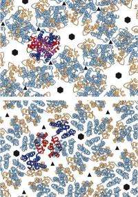
As a retrovirus matures, the two parts of its shell protein (red and blue or yellow and blue) dramatically rearrange themselves, twisting and moving away from each other. Credit: EMBL/T.Bharat
Scientists at the European Molecular Biology Laboratory (EMBL) in Heidelberg, Germany, have for the first time uncovered the detailed structure of the shell that surrounds the genetic material of retroviruses, such as HIV, at a crucial and potentially vulnerable stage in their life cycle: when they are still being formed. The study, published online today in Nature, provides information on a part of the virus that may be a potential future drug target.
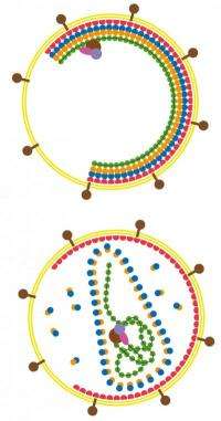
Retroviruses essentially consist of genetic material encased in a protein shell, which is in turn surrounded by a membrane. After entering a target cell – in the case of HIV, one of the cells in our immune system – the virus replicates, producing more copies of itself, each of which has to be assembled from a medley of viral and cellular components into an immature virus. "All the necessary components are brought together within the host cell to form the immature virus, which then has to mature into a particle that's able to infect other cells" says John Briggs, who led the research at EMBL. "We found that when it does, the changes to the virus' shell are more dramatic than expected."
The role and shape of the protein shell (blue/orange) changes from the immature (top) to the mature form of the virus (bottom). Credit: EMBL/T.Bharat
Both the mature and immature virus shells are honeycomb-like lattices of hexagon-shaped units. Using a combination of electron microscopy and computer-based methods, Briggs and colleagues investigated which parts of the key proteins stick together to build the honeycomb of the immature shell. These turned out to be very different from the parts that build the mature shell. This knowledge will help scientists to unravel how the immature virus is assembled in the cell and how the shell proteins rearrange themselves to go from one form to the other.
As a retrovirus matures, the two parts of its shell protein (red and blue or yellow and blue) dramatically rearrange themselves, twisting and moving away from each other. Credit: EMBL/T.Bharat
Findings such as these may one day prove valuable to those wanting to design new types of anti-retroviral therapies. Many anti-retroviral drugs already block the enzyme that would normally separate components of the immature shell to allow it to mature. But there are currently no approved drugs that act on that shell itself and prevent the enzyme from locking on.
Although the virus shells imaged in this study were derived from the Mason-Pfizer monkey virus and made artificially in the laboratory, they closely resemble those of both this virus and HIV – which are very similar – in their natural forms.
"We still need a lot more detailed information before drug design can really be contemplated," Briggs concludes, "but finally being able to compare mature and immature structures is a step forwards."
Journal information: Nature
Provided by European Molecular Biology Laboratory


