Studying slimy substances for a cleaner environment
Extracellular polymeric substances, or EPS, are the slimy material that bacteria excrete and surround themselves with as they form biofilms. EPS are mostly water (up to 95%), but the remaining ingredients include a complex mixture of lipids, saccharides (sugars), and bacterial debris such as cell fractions, DNA, and proteins. Because of the crucial role microbial communities can play in the health of both humans and ecosystems, EMSL scientist Alice Dohnalkova has been working with a team of researchers to uncover new discoveries about EPS and their unknown role in governing molecular interactions.
“In recent years, we have learned that EPS are so much more than just the sticky material that holds the bacterial communities in the biofilm together,” Dohnalkova said. “They also contain macromolecular complexes capable of metal reduction.”
BACTERIA FIGHTING THE GOOD FIGHT
Metal reduction is a chemical process with high-stakes environmental implications. Heavy metals and radionuclides in contaminated soils pose a very serious problem in terms of their future transport and fate in the subsurface, including potential groundwater contamination. Some soil bacteria have the ability to carry out electron transport functions that reduce these metals into their less soluble forms, and thus prevent their spread in the subsurface. Understanding to what extent microbes can be used as “good guys” in this way is critical for remediation of contaminated sites around the world. For example, uranium contamination at the Department of Energy’s Hanford Nuclear Reservation could be alleviated using such “bioremediation” efforts as part of the overall cleanup strategy.
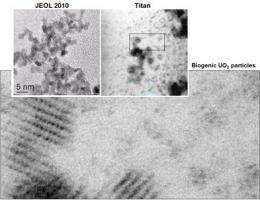
However, microbial metal reduction is a complex process. So, Dohnalkova and collaborators from Pennsylvania State University, the South Dakota School of Mines and Engineering, and Pacific Northwest National Laboratory are leveraging their research ideas using state-of-the-art instrumentation available in EMSL. A new high-resolution Titan scanning/transmission electron microscope (STEM) enables angstrom-resolution imaging, offering unprecedented views of the newly formed biominerals at the first stages of particle nucleation. Understanding these underlying mechanisms requires observations at the smallest scale available. For perspective, a strand of human hair is about a half million angstroms thick.
“These nanoparticles were previously calculated using theory and modeled based on previous X-ray methods,” Dohnalkova said. “But we were never able to provide the direct evidence of their presence—until we got a chance to image them with the new atomic-resolution STEM.”
THE ANCHOR INSTRUMENT
EMSL’s Titan STEM, manufactured by FEI and acquired through funding from the American Recovery and Reinvestment Act, is a high-demand instrument for researchers like Dohnalkova who seek atomic-level images to support high-impact discoveries. It is referred to as a “multi-purpose tool” for research involving energy materials, catalysis, interfacial phenomena, and nanostructured materials. The Titan and its various features and attachments enable studies of dynamic scientific processes in realistic environments, in part by using sample holders that can control temperature, electrical current, and gaseous environment. Combined with world-class sample preparation and located in EMSL’s new Quiet Wing—a facility that minimizes acoustic interferences, vibrations, and magnetic field to optimize instrument performance—the Titan is enabling science not previously possible in many research areas, including Dohnalkova’s work with EPS.
“In some ways, it’s the anchor of our suite of capabilities,” said Bruce Arey, an EMSL technologist in the user facility’s microscopy group.
Like the anchor sprinter on an Olympic relay team, the Titan cannot produce results on its own. EMSL recognized the microscope must be customized and integrated with other instruments, techniques, and expertise in a unique environment. When these legs of the race are run correctly, EMSL users can allow the Titan to “take the baton” for world-record-setting results.
A PIECE OF THE PUZZLE FOR ORGANIC CLEANUP
In the case of studying bacterial EPS to support uranium reduction through bioremediation, Dohnalkova and her collaborators have teamed up to correlate a variety of methods. As a result, they provide a more complete view of the microbial-metal interactions and how they are facilitated by EPS. Molecular biology, kinetic studies, crystallography, and chemical imaging are just a few areas of interest for the multidisciplinary, multi-institutional team. According to Dohnalkova, strong communication among the team and scientific leadership from PNNL’s Microbial Communities Initiative has led to better results.
“For the first time, we were able to visualize newly formed nanocrystalline uranium (IV) oxide particles that were transformed by bacteria from a solution of uranium (VI),” Dohnalkova said, describing the team’s most intriguing recent findings. “The existence of these tiny particles—sized on the order of single angstroms—was previously predicted by other means, but the direct evidence was only possible by high-resolution imaging using the Titan.
“This particular data is a significant puzzle piece that fits into the larger picture of microbial interaction with the environmental contaminants.”
Being able to finally see the metal-reducing transformation and the mechanisms causing it helps refine the complex, predictive science of bioremediation. At the same time, it energizes Dohnalkova to continue pushing for further discoveries.
“I’m excited to see a great story developing,” she said. “I believe the concept of the ‘good bacteria’ is attractive to all environmentally conscious people. DOE also strongly supports other closely related bioremediation research efforts on, for example, bacteria dealing with oil spills.
“As we grow our understanding, bacterial remediation is continuing to prove itself as an elegant, ‘organic’ way to protect our environment from these pollutants.”
The research team is sharing their results with the scientific community via publications and conference presentations.
More information: Dohnalkova AC, et al. 2011. “Imaging Hydrated Microbial Extracellular Polymers: Comparative Analysis by Electron Microscopy.” Applied and Environmental Microbiology 77(4):1254-1262. DOI: 10.1128/AEM.02001-10
Marshall MJ, et al. 2008. “Hydrogenase- and outer membrane c-type cytochrome-facilitated reduction of technetium(VII) by Shewanella oneidensis MR-1.” Environmental Microbiology 10(1):125-136. DOI: 10.1111/j.1462-2920.2007.01438.x
Burgos WD, et al. 2008. “Characterization of uraninite nanoparticles produced by Shewanella oneidensis MR-1.” Geochimica et Cosmochimica Acta 72(20):4901-4915. DOI: 10.1016/j.gca.2008.07.016
Sani RK, et al. 2008. “Comparison of uranium(VI) removal by Shewanella oneidensis MR-1 in flow and batch reactors.” Water Research 42(12):2993-3002. DOI: 10.1016/j.watres.2008.04.003
Senko JM, et al. 2007. “The effect of U(VI) bioreduction kinetics on subsequent reoxidation of biogenic U(IV).” Geochimica et Cosmochimica Acta 71(19):4644-4654. DOI: 10.1016/j.gca.2007.07.021
Provided by Environmental Molecular Sciences Laboratory


