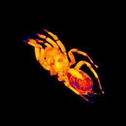
Scientists can color the MRI images to highlight organs. The heart is shown in lighter colors in the posterior part of the body. Credit: Gavin Merrifield
Researchers have used a specialised Magnetic Resonance Imaging (MRI) scanner on tarantulas for the first time, giving unprecedented videos of a tarantula's heart beating.
"In the videos you can see the blood flowing through the heart and tantalisingly it looks as though there might be 'double beating' occurring, a distinct type of contraction which has never been considered before. This shows the extra value of using a non-invasive technique like MRI," says PhD researcher Gavin Merrifield who is presenting the research at the Society for Experimental Biology Annual Conference in Glasgow on the 1st July 2011.
Researchers from Edinburgh University used MRI scanners at the Glasgow Experimental MRI centre that had been built for medical research on rodents to produce images of a tarantula's heart and gut. The use of MRI reduces the need for dissection and provides greater insight to internal workings as the animals are live and unharmed.
This shows MRI images of a tarantula’s heart beating in real time. Credit: Gavin Merrifield
Heart rate and cardiac output were measured, giving much more accurate readings than previous methods which were either indirect or highly invasive.
Application of this technology is often purely medical; however diversifying its use can have practical benefits or even answer fundamental biological questions. "One potential practical use of this research is to ascertain the chemical composition of spider venom," says Mr. Merrifield. "Venom has applications in agriculture as a potential natural pesticide. On the more academic side of things if we can link MRI brain scans with a spider's behaviour, and combine this with similar data from vertebrates, we may clarify how intelligence evolved."
Provided by Society for Experimental Biology


