Moving microscopic vision into another new dimension

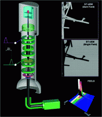
Scientists who pioneered a revolutionary 3-D microscope technique are now describing an extension of that technology into a new dimension that promises sweeping applications in medicine, biological research, and development of new electronic devices. Their reports on so-called 4-D scanning ultrafast electron microscopy, and a related technique, appear in two papers in the Journal of the American Chemical Society.
Chemistry Nobel Laureate Ahmed H. Zewail and colleagues moved high-resolution images of vanishingly small nanoscale objects from three dimensions to four dimensions when they discovered a way to integrate time into traditional electron microscopy observations. Their laser-driven technology allowed researchers to visualize 3-D structures such as a ring-shaped carbon nanotube while it wiggled in response to heating, over a time scale of femtoseconds. A femtosecond is one millionth of one billionth of a second. But the 3-D information obtained with that approach was limited because it showed objects as stationary, rather than while undergoing their natural movements.
The scientists describe how 4-D scanning ultrafast electron microscopy and scanning transmission ultrafast electron microscopy overcome that limitation, and allow deeper insights into the innermost structure of materials. The reports show how the technique can be used to investigate atomic-scale dynamics on metal surfaces, and watch the vibrations of a single silver nanowire and a gold nanoparticle. The new techniques "promise to have wide ranging applications in materials science and in single-particle biological imaging," they write.
More information: “4D Scanning Ultrafast Electron Microscopy: Visualization of Materials Surface Dynamics”, J. Am. Chem. Soc., Article ASAP. DOI:10.1021/ja203821y
Abstract
We report the development of 4D scanning transmission ultrafast electron microscopy (ST-UEM). The method was demonstrated in the imaging of silver nanowires and gold nanoparticles. For the wire, the mechanical motion and shape morphological dynamics were imaged, and from the images we obtained the resonance frequency and the dephasing time of the motion. Moreover, we demonstrate here the simultaneous acquisition of dark-field images and electron energy loss spectra from a single gold nanoparticle, which is not possible with conventional methods. The local probing capabilities of ST-UEM open new avenues for probing dynamic processes, from single isolated to embedded nanostructures, without being affected by the heterogeneous processes of ensemble-averaged dynamics. Such methodology promises to have wide-ranging applications in materials science and in single-particle biological imaging.
Provided by American Chemical Society


