Human cells filmed instantly messaging for first time
Cells tugged in one direction sent biochemical signals in the opposite direction in the form of a signature pattern of fluorescent light
Researchers at UCSD and UC Irvine have captured on video for the first time chemical signals that traverse human cells in response to tiny mechanical jabs, like waves spreading from pebbles tossed into a pond. The scientists released the videos and technical details that explain how the visualization effect was created as part of a paper published in the April 21 issue of Nature.
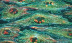
Image: Not only did researchers at UCSD and UCI visualize a mechanically induced signal traverse human cells, but they also showed that actin filaments and microtubules are involved in the process.
The researchers working at the UCSD Jacobs School of Engineering's Department of Bioengineering developed a novel molecular "reporter" system, which allowed the dynamic visualization of the activation of an important protein called Src. Peter Yingxiao Wang, lead author of the paper and a post-doctoral researcher in UCSD's Jacobs School of Engineering spent two years designing the reporter molecules to light up selectively only when Src was activated, and not other proteins.
Wang and his co-workers first demonstrated that the novel system was effective in visualizing Src activation in response to a known chemical stimulant, epidermal growth factor. Next, they studied the effect of mechanical stimuli on Src activation. Using technology developed at the Beckman Laser Institute at UC Irvine by its founding director Michael Berns, Wang and Elliott Botvinick, a postdoctoral researcher at UCSD Department of Bioengineering and the Beckman Laser Institute at UC Irvine, attached small, sticky beads to cells and gently tugged the beads to and fro with laser power acting as invisible "tweezers." As the laser tweezers moved the beads in one direction, a video camera attached to a specially equipped microscope recorded the dynamic movement of biochemical signals in the opposite direction in the form of a signature pattern of fluorescent light. The fine spatial and temporal resolution was made possible by a technology called fluorescence resonance energy transfer.
"We had no idea what to expect," said Wang. "The first time we saw these incredible waves spreading across the cells I just said 'Whoa, this is amazing.' We expected to see a signal where the tweezers were pulling the beads, but we did not envision such a directional wave propagating away from the beads." Wang worked on this project under the joint advisorship of Shu Chien, a professor of bioengineering and medicine and director of the Whitaker Institute of Biomedical Engineering at UCSD, and Roger Y. Tsien, professor of pharmacology, chemistry, and biochemistry and investigator with the Howard Hughes Medical Institute at UCSD.
Src is one of a large group of enzymes called kinases that attach a phosphate molecule to one or more target proteins in the cell. This phosphorylation reaction typically switches the target protein from inactive to active status. Many diseases can result either when a kinase gene is mutated and can't properly phosphorylate its targets, or when a normal kinase becomes overactive or not sufficiently active. Indeed, Src has been shown to play a key role in cell growth and development, and in the genesis of cancer, atherosclerosis, and many other disease conditions.
"This study amounts to a proof of principle that if we can visualize the activation of one kinase, we can do the same for many others using the same approach," said Chien, the senior author of the paper. Not only are those additional studies expected to reveal temporal and spatial patterns of kinase activation, but Chien also predicted that there will be practical spin offs.
For example, cells usually tightly control the activity of Src, but in certain cancers its activity is abnormality high. "We think that our ability to measure Src activity with this new visualization technique would be useful as a diagnostic test for many cancers," said Chien. The William J. von Liebig Center for Entrepreneurism and Technology Advancement at UCSD's Jacobs School has provided Chien and Wang with funding to commercialize the new visualization technology as a cancer-detection tool.
The researchers showed that actin filaments and microtubules, structural elements that traverse cells like the ribs of an umbrella, could function as conduits for the spread of biochemical signals. Indeed, when Wang disrupted either actin filaments or microtubules in his test cells, the activation signal no longer spread across the cell. These results suggest that the activation of Src traverses these filamentous structures.
Source: University of California - San Diego


