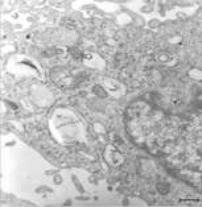
Role of autophagic and lysosomal pathways in ischemic brain injury

Previous studies by Shaohua Gu and team from Shanghai Pudong New Area Zhoupu Hospital showed that rapamycin-induced autophagy decreased the rate of apoptosis, but the rate of apoptosis was increased after the autophagy inhibitor, 3-methyladenine, was used, indicating autophagy may be involved in mediating neuronal death in cerebral ischemia.
A recent study reported by Gu et al showed that autophagic and lysosomal activity is increased in ischemic neurons, and the activation of autophagic and lysosomal pathways can provide nutrition and energy for the survival of ischemic neurons, which was published in the Neural Regeneration Research (Vol. 8, No. 23, 2013). Researchers believe that abnormal components in cells can be eliminated through upregulating cell autophagy or inhibiting autophagy after ischemic brain injury, resulting in a dynamic balance of substances in cells. Moreover, drugs that interfere with autophagy may be potential therapies for the treatment of brain injury.
More information:
Gu ZH, Sun YY, Liu KY, Wang F, Zhang T, Li Q, Shen LW, Zhou L, Dong L, Shi N, Zhang Q, Zhang W, Zhao MZ, Sun XJ. The role of autophagic and lysosomal pathways in ischemic brain injury. Neural Regen Res. 2013;8(23):2117-2125.
Provided by Neural Regeneration Research