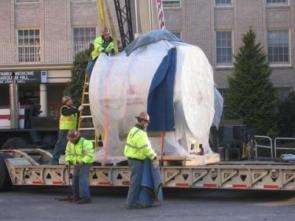
OHSU: 'Two-of-a-kind' MRI magnet arrived

As OHSU's new Advanced Imaging Research Center (AIRC) continues its expansion, the major component of the final, and most unique instrument for the world-class facility has arrived. On Saturday, April 22, the AIRC took delivery of a 12Tesla (T) magnet, the centerpiece of a rare and cutting-edge MRI system.
"This state-of-the-art 12T system will provide some of the clearest, and likely some of the most useful, images in the history of MRI science," explained Charles Springer, Ph.D., AIRC director and professor of physiology and pharmacology in the OHSU School of Medicine. Springer also serves as a professor of biomedical engineering at OHSU and is a member of the OHSU Cancer Institute. His arrival, along with the establishment of the AIRC, took place through the Oregon Opportunity, a public-private partnership to increase the health and economic benefits of OHSU research for all Oregonians.
The 12T instrument with a magnetic field 120,000 times stronger than that of the Earth was purchased with assistance from a $1.75 million grant provided by the W. M. Keck Foundation. This is the third MRI magnet delivery for OHSU this year. In early January, a 3T magnet was lifted by crane into OHSU's new Biomedical Research Building, the primary home of the AIRC. In late January, OHSU received delivery of a 60 ton, 7T magnet. The extreme size and weight of this piece of equipment, paired with its fragility, resulted in a precise initial positioning that lasted three days. By comparison, the 12T magnet weighs approximately 12 tons (24,000 lbs). However, the magnetic field that will be generated is one of the strongest in the world for a magnet of this relatively large physical size. The inner diameter of its horizontal cylindrical bore is 31 centimeters. In fact, the National Institutes of Health in Bethesda, Md. -- the base of operations for federally funded health research -- is currently the only other site in the world for a 12T MRI system of this size. Both of OHSU's 12T and 7T systems will be housed in the W. M. Keck Foundation High-Field Laboratory within the AIRC.
"It will be exciting to watch the wonderful science that is sure to emerge from such an outstanding team of researchers doing experiments with this very unique 12T MR system," said Michael Garwood, Ph.D. Who serves as the associate director of the Center for Magnetic Resonance Research at the University of Minnesota, a national leader in imaging technology and expertise. "The possibility to conduct experiments at 12T will certainly lead to important discoveries and to an expansion of MRI's role in assessing functional and molecular properties of diseases non-invasively."
Once in operation, the 12T will be used for a variety of human health studies in rodents including research into neurological disorders such as multiple sclerosis, Alzheimer's and stroke. For instance, William Rooney, Ph.D., a staff scientist at the AIRC will measure water movement in the brain. These studies will assist in a greater understanding of the development of brain tumors and diseases like MS.
"Using a dyeing agent, lower field instruments have allowed us to witness the progression of MS," explained Rooney. "Specifically, we've been able to see the development of brain lesions associated with disease. With a cutting-edge, high-field MRI system, we will both literally and figuratively get a much clearer picture of what is going on in the brain. This technology will provide us with a powerful noninvasive window into the brain and allow us to explore unique aspects of disease."
Research conducted by OHSU's internationally recognized Blood-Brain Barrier Program will also greatly benefit by the arrival of the 12T magnet. The blood-brain barrier is a natural protector that prevents toxins from circulating into the brain. The barrier also plays a role in the delicate balance of pressure in the brain. A better understanding of the barrier is necessary for improving chemotherapy for brain tumors. In addition, it is believed that the barrier has a significant role in neurological diseases such as MS.
The director of OHSU's Blood Brain Barrier Program, Edward Neuwelt, M.D., and his colleagues have also designed new methods for highlighting white blood cells in the brain so that they may be seen by MRI. Given that white blood cells play a very important role in the body's immune system, gaining new knowledge about their impacts on the brain could be significant in a variety of ways.
Springer and his colleagues will be involved in many of these studies, including research aimed at gaining new understanding of stroke in studies conducted using the 12T magnet. Substance and alcohol abuse studies by OHSU researchers will also be greatly aided by the addition of the 12T system.
MRI Technology:
Magnetic resonance imaging is a technique developed to create high-resolution 3-D pictures of any location in the body. MRI uses computer-controlled radio frequency (RF) waves and magnetic field gradients inside large magnets strong enough to generate fields tens of thousands of times stronger than the earth's magnetic field at its surface. The combination of the RF waves and this magnetic field causes the hydrogen nuclei of water and other molecules in the body to respond. RF signals from these responses can then be detected and used to create 3-D images. Unlike X-ray imaging or CAT (computed axial tomorography) scanning, MRI does not employ ionizing radiation and poses no inherent health risks to subjects. Magnetic field strengths are measured in Teslas. (One Tesla represents a strength approximately 10,000 times that of the earth's magnetic field.)
For instance, an MRI instrument with a 1.5T magnet can potentially provide a superior image of the body region under study than one with a 1T magnet. The main reason for this is that the strengths of the tissue water-hydrogen RF signals increase with increasing magnetic field strength. Prior to the arrivals of the AIRC high-field magnets, OHSU had two 1.5T and two 3T MRI units for use in clinical diagnosis. The Portland VA Medical Center on Marquam Hill also has a 1.5T MRI scanner.
Source: Oregon Health & Science University


