Picturing pain could help unlock its mysteries and lead to better treatments
Understanding the science behind pain, from a simple "ouch" to the chronic and excruciating, has been an elusive goal for centuries. But now, researchers are reporting a promising step toward studying pain in action. In a study published in the Journal of the American Chemical Society, scientists describe the development of a new technique, which they tested in rats, that could result in better ways to relieve pain and monitor healing.
Sandip Biswal, Frederick T. Chin, Justin Du Bois and colleagues note that current ways to diagnose pain basically involve asking the patient if something hurts. These subjective approaches are fraught with bias and can lead doctors in the wrong direction if a patient doesn't want to talk about the pain or can't communicate well. It can also be difficult to tell how well a treatment is really working. No existing method can measure pain intensity objectively or help physicians pinpoint the exact location of the pain. Past research has shown an association between pain and a certain kind of protein, called a sodium channel, that helps nerve cells transmit pain and other sensations to the brain. Certain forms of this channel are overproduced at the site of an injury, so the team set out to develop an imaging method to visualize high concentrations of this protein.
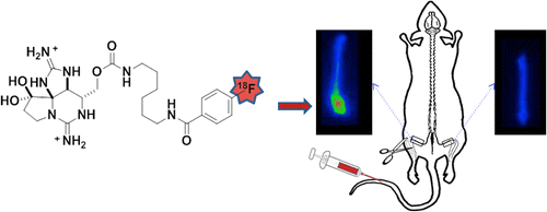
They turned to a small molecule called saxitoxin, produced naturally by certain types of microscopic marine creatures, and attached a signal to it so they could trace it by PET imaging. PET scanners are used in hospitals to diagnose diseases and injuries. When the researchers injected the molecule into rats, often a stand-in for humans in lab tests, they saw that the molecule accumulated where the rats had nerve damage. The rats didn't show signs of toxic side effects. The work is one of the first attempts to mark these sodium channels in a living animal, they say.
More information: "A 18F-Labeled Saxitoxin Derivative for in Vivo PET-MR Imaging of Voltage-Gated Sodium Channel Expression Following Nerve Injury" J. Am. Chem. Soc., 2013, 135 (48), pp 18012–18015. DOI: 10.1021/ja408300e
Abstract
Both chronic and neuropathic pain conditions are associated with increased expression of certain voltage-gated sodium ion channel (NaV) isoforms in peripheral sensory neurons. A method for noninvasive imaging of these channels could represent a powerful tool for investigating aberrant expression of NaV and its role in pain pathogenesis. Herein, we describe the synthesis and evaluation of a positron emission tomography (PET) radiotracer targeting NaVs, the design of which is based on the potent, NaV-selective inhibitor saxitoxin. Both autoradiography analysis of sciatic nerves excised from injured rats as well as whole animal PET-MR imaging demonstrate that a systemically administered [18F]-labeled saxitoxin derivative concentrates at the site of nerve injury, consistent with upregulated sodium channel expression following axotomy. This type of PET agent has potential use for serial monitoring of channel expression levels at injured nerves throughout wound healing and/or following drug treatment. Such information may be correlated with pain behavioral analyses to help shed light on the complex molecular processes that underlie pain sensation.
Journal information: Journal of the American Chemical Society
Provided by American Chemical Society


