How the brain folds to fit
During fetal development of the mammalian brain, the cerebral cortex undergoes a marked expansion in surface area in some species, which is accommodated by folding of the tissue in species with most expanded neuron numbers and surface area. Researchers have now identified a key regulator of this crucial process.
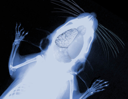
Different regions of the mammalian brain are devoted to the performance of specific tasks. This in turn imposes particular demands on their development and structural organization. In the vertebrate forebrain, for instance, the cerebral cortex – which is responsible for cognitive functions – is remarkably expanded and extensively folded exclusively in mammalian species. The greater the degree of folding and the more furrows present, the larger is the surface area available for reception and processing of neural information. In humans, the exterior of the developing brain remains smooth until about the sixth month of gestation. Only then do superficial folds begin to appear and ultimately dominate the entire brain in humans. Conversely mice, for example, have a much smaller and smooth cerebral cortex.
"The mechanisms that control the expansion and folding of the brain during fetal development have so far been mysterious," says Professor Magdalena Götz, a professor at the Institute of Physiology at LMU and Director of the Institute for Stem Cell Research at the Helmholtz Center Munich. Götz and her team have now pinpointed a major player involved in the molecular process that drives cortical expansion in the mouse. They were able to show that a novel nuclear protein called Trnp1 triggers the enormous increase in the numbers of nerve cells which forces the cortex to undergo a complex series of folds. Indeed, although the normal mouse brain has a smooth appearance, dynamic regulation of Trnp1 results in activating all necessary processes for the formation of a much enlarged and folded cerebral cortex. Levels of Trnp1 control expansion and folding "Trnp1 is critical for the expansion and folding of the cerebral cortex, and its expression level is dynamically controlled during development," says Götz. In the early embryo, Trnp1 is locally expressed in high concentrations. This promotes the proliferation of self-renewing multipotent neural stem cells and supports tangential expansion of the cerebral cortex. The subsequent fall in levels of Trnp1 is associated with an increase in the numbers of various intermediate progenitors and basal radial glial cells. This results in the ordered formation and migration of a much enlarged number of neurons forming folds in the growing cortex.
The findings are particularly striking because they imply that the same molecule – Trnp1 – controls both the expansion and the folding of the cerebral cortex and is even sufficient to induce folding in a normally smooth cerebral cortex. Trnp1 therefore serves as an ideal starting point from which to dissect the complex network of cellular and molecular interactions that underpin the whole process. Götz and her colleagues are now embarking on the next step in this exciting journey - determination of the molecular function of this novel nuclear protein Trnp1 and how it is regulated.
More information: Trnp1 regulates expansion and folding of the mammalian cerebral cortex by control of radial glial fate, Ronny Stahl, Tessa Walcher, Camino De Juan Romero, Gregor Alexander Pilz, Silvia Cappello, Martin Irmler, José Miguel Sanz Anquela, Johannes Beckers, Robert Blum, Víctor Borrell, and Magdalena Götz, Cell 2013, doi: 10.1016/j.cell.2013.03.027


