Diagnostic brain tumor test could revolutionize care of patients

Researchers at UT Southwestern Medical Center have developed what they believe to be the first clinical application of a new imaging technique to diagnose brain tumors. The unique test could preclude the need for surgery in patients whose tumors are located in areas of the brain too dangerous to biopsy.
This new magnetic resonance spectroscopy (MRS) technique provides a definitive diagnosis of cancer based on imaging of a protein associated with a mutated gene found in 80 percent of low- and intermediate-grade gliomas. Presence of the mutation also means a better prognosis.
"To our knowledge, this is the only direct metabolic consequence of a genetic mutation in a cancer cell that can be identified through noninvasive imaging," said Dr. Elizabeth Maher, associate professor of internal medicine and neurology at UT Southwestern and senior author of the study, available online in Nature Medicine. "This is a major breakthrough for brain tumor patients."
UT Southwestern researchers developed the test by modifying the settings of a magnetic resonance imaging (MRI) scanner to track the protein's levels. The data acquisition and analysis procedure was developed by study lead author Dr. Changho Choi, associate professor of the Advanced Imaging Research Center (AIRC) and radiology. Previous research linked high levels of this protein to the mutation, and UT Southwestern researchers already had been working on MRS of gliomas to find tumor biomarkers.
"Our next step is to make this testing procedure widely available as part of routine MRIs for brain tumors. It doesn't require any injections or special equipment," said Dr. Maher, medical director of UT Southwestern's neuro-oncology program.
To substantiate the test as a diagnostic tool, biopsy samples from 30 glioma patients enrolled in the UT Southwestern clinical trial were analyzed; half had the mutation and expected high levels of the protein. MRS imaging of these patients had been done before surgery and predicted, with 100 percent accuracy, which patients had the mutation.
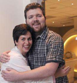
For Thomas Smith of Grand Prairie, the test helped determine the best time to begin chemotherapy. When an MRS scan showed a sharp rise in the 25-year-old's protein levels, this indicated to his health care team that his tumor was moving from dormancy to rapid growth.
"We treated him with chemotherapy and his protein levels came down," Dr. Maher said.
Before participating in the study, Mr. Smith had tumor removal surgery in 2007. Because part of the tumor could not be safely removed, however, he continued to suffer seizures and had other neurological problems. Since chemotherapy, his symptoms have diminished.
"I did six rounds of chemo, every six weeks," Mr. Smith said. "My seizures stopped and all my symptoms improved. I am only on anti-seizure medication now."


